Fluid Volume Disturbance

Fluid Volume Deficit (Hypovolemia)

 Definition:fluid volume deficit (FVD) occurs when loss of extracellular fluid volume exceeds the intake of fluid

It occurs when water and electrolyte are lost in the same proportion as they exist in normal fluids Fluid volume deficit (hypovolemia) should not be confused with the term dehydration, which refers to loss of water alone with increased serum sodium level, FVD may occur alone or in combination with other imbalances

Pathophysiology 

FVD results from loss of body fluids and occurs more rapidly when coupled with decreased fluid intake

FVD can develop from inadequate intake alone if the decreased intake is prolonged

Causes of FVD include abnormal fluid losses such as resulting from vomiting, diarrhea, G.I suctioning and sweating and decreased intake as in Nausea or irritability to gain access to fluids (Beck – 2000) Additional risk factors diabetes inspidius, adrenal insufficiency, osmotic dieresis hemorrhage and coma

Clinical Manifestation 

Fluid volume deficit can develop rapidly and can be

  • Mild
  • Moderate
  • Or severe depending on the degree of fluid loss

Important characteristics of FVD include

  • Acute wt loss
  • Decreased skin turgor
  • Oliguria
  • Concentrated urine
  • Postural hypotension
  • A weak, rapid heart rate
  • Flattened neck veins
  • Increased temperature
  • Decreased central venous pressure
  • Cool
  • Clamming skin related to peripheral vasoconstriction
  • Thirst
  • Anorexia
  • Nausea
  • Muscles weakness
  • Cramps

Diagnosis

  • BUN (blood urine nitrogen) related to serum cretinine concentration (a ratio greater than 20:1)
  • Health history
  • Physical examination
  • Serum electrolytes changes serum K, Na (hypokalemia, hyponatermia or hypernatermia, hyperkalemia)
  • Urine specific gravity increased

Medical Management 

  • Planning of correction of fluid loss for patient with fluid volume deficit (FVD)
  • The health care provider consider the usual maintenance requirements of the patient and other factors (such as fever) that can influence fluid needs
  • When deficit is not severe the oral route is preferred
  • In acute and severe losses the I/V route is required isotonic electrolyte solutions e.g lactated Ringer’s or 0.9% N/S are frequently used to treat hypotension pts with FVD
  • Maintain and assess I/O (intake and output chart)
  • Wt: , vital signs, central venous pressure, level of consciousness (LOC), breath sounds, skin color
  • The rate of fluid administration is based on severity of loss of and patient hemodynamic response

Nursing Management 

  • To assess for fluid volume deficit
  • Nurse monitors and measures fluid intake and output at least every 8 hours and sometime hourly
  • Vital signs closely monitored
  • Nurse should observe for weak pulse and postural hypotension
  • Skin and tongue turgor is monitored on regular basis
  • Preventing FVD – as diarrhea
  • Correcting FVD – if patient is unable to take fluid the orally the nurse should give fluid parental I/V

Manifestations Of Urinary System Disorders


MANIFESTATIONS OF URINARY SYSTEM DISORDERS
General Manifestations Specific Manifestations Related to Urinary System
 Edema  Pain  Patterns of Urination  Urine Output  Urine Composition
Fatigue Facial (periorbital) Dysuria Frequency Anuria Concentrated
Headaches Ankle Flank or Urgency Oliguria Dilute
Blurred vision Ascites costovertebral Hesitancy of stream Polyuria Hematuria
Elevated blood pressure Anasarca angle Change in stream Pyuria
Anorexia Sacral Groin Retention Color (red, brown,
Nausea and vomiting Suprapubic Dysuria yellowish green)
Chills Nocturia
Itching Incontinence
Excessive thirst Stress incontinence
Change in body weight Dribbling
Cognitive changes

Urinary System

HEALTH HISTORY
Health Perception–Health Management
• How is your energy level compared with 1 yr ago?
• Do you notice any visual changes?*
• Have you ever smoked? If yes, how many packs per day?
Nutritional-Metabolic
• How is your appetite?
• Has your weight changed over the past yr?*
• Do you take vitamins, herbs, or any other supplements?*
• How much and what kinds of fluids do you drink daily?
• How many dairy products and how much meat do you eat?
• Do you drink coffee? Colas? Tea?
• Do you eat chocolate?
• Do you spice your food heavily?*
Elimination
• Are you able to sit through a 2-hr meeting or ride in a car for 2 hr without urinating?
• Do you awaken at night with the desire to urinate? If so, how many times does this occur during an average night?
• Do you ever notice blood in your urine?* If so, at what point in the urination does it occur?
• Do you ever pass urine when you do not intend to? When?
• Do you use special devices or supplies for urine elimination or control?*
• How often do you move your bowels?
• Do you ever experience constipation?
• Do you frequently experience diarrhea? Do you ever have problems controlling your bowels? If so, do you have problems controlling the passage of gas? Watery or liquid stool? Solid stool?
Activity-Exercise
• Have you noticed any changes in your ability to do your usual daily activities?*
• Do certain activities aggravate your urinary problem?*
• Has your urinary problem caused you to alter or stop any activity or exercise?*
• Do you require assistance in moving or getting to the bathroom?*
Sleep-Rest
• Do you awaken at night from an urge to urinate?*
• Do you awaken at night from pain or other problems and urinate as a matter of routine before returning to sleep?*
• Do you experience daytime sleepiness and fatigue as a result of nighttime urination?*
Cognitive-Perceptual
• Do you ever have pain when you urinate?* If so, where is the pain?
Self-Perception–Self-Concept
• How does your urinary problem make you feel about yourself?
• Do you perceive your body differently since you have developed a urinary problem?
Role-Relationship
• Does your urinary problem interfere with your relationships with family or friends?*
• Has your urinary problem caused a change in your job status or
affected your ability to carry out job-related responsibilities?*
Sexuality-Reproductive
• Has your urinary problem caused any change in your sexual pleasure or performance?*
• Do you have hygiene problems related to sexual activities that cause you concern?*
Coping–Stress Tolerance
• Do you feel able to manage the problems associated with your urinary problem? If not, explain.
• What strategies are you using to cope with your urinary problem?
Values-Beliefs
• Has your present illness affected your belief system?*
• Are your treatment decisions related to your urinary problem in conflict with your value system?*

Hypertension

Key facts

  • Hypertension, or high blood pressure, is a life-threatening medical condition that greatly raises the chances of cardiovascular disease, stroke, dementia, and renal failure, and many others.
  • It is estimated that 1.28 billion individuals aged 30-79 years worldwide have hypertension, with the majority (two-thirds) living in low- and middle-income countries.
  • Additionally, it is estimated that 46 percent of adults with hypertension do not know they have the illness.
  • Hypertension is detected in less than half of adult patients (42%).
  • Only about a fifth of persons with hypertension (21%) have it under control.
  • Premature mortality due to hypertension is a serious global health problem.

What exactly is hypertension?

Blood pressure is the force produced by flowing blood on the arterial walls, the body’s primary blood vessels. Hypertension is too high blood pressure.

Blood pressure is expressed as a pair of numbers. The first number (systolic) shows the blood vessel pressure when the heart contracts or beats. The second number (diastolic) indicates the pressure in the blood arteries between heartbeats.

When tested on two separate days, hypertension is diagnosed if the systolic blood pressure readings on both days are 140 mmHg and/or the diastolic blood pressure readings on both days are 90 mmHg.

Know the Risk Factors for High Blood Pressure (Hypertension) | Top 10 Home  Remedies
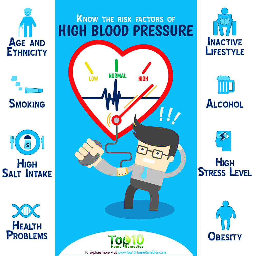
What risk factors are associated with hypertension?

Unhealthy diets (excessive salt consumption, a diet heavy in saturated fat and trans fats, and a poor intake of fruits and vegetables), physical inactivity, cigarette and alcohol use, and being overweight or obese are modifiable risk factors.

Non-modifiable risk factors include a family history of hypertension, age over 65, and coexisting conditions like diabetes or renal illness.

Pathophysiology Of Primary Hypertension

Hypertension is characterised by a consistently elevated SVR.

Retention of water and sodium: A high sodium intake may activate many pressor systems and produce water retention.

High plasma renin activity (PRA) causes an increase in the conversion of angiotensinogen to angiotensin I, which causes arteriolar constriction, vascular hypertrophy, and aldosterone production.

Stress and increased SNS activity: Anger, fear, and pain have an effect on arterial pressure. Normally protective physiological reactions to stress may continue to a pathologic degree, resulting in a protracted increase in SNS activity. Enhanced SNS activation results in increased vasoconstriction, heart rate, and renin release.

Insulin resistance and hyperinsulinemia are frequent in primary hypertension, as are abnormalities of glucose, insulin, and lipoprotein metabolism. Vascular hypertrophy and enhanced renal salt reabsorption are supplementary pressor effects of insulin.

Endothelial cell dysfunction is characterised by a diminished vasodilator response to nitric oxide in some hypertensive individuals. Nitric oxide, an endothelium-derived relaxing factor (EDRF), helps maintain low arterial tone at rest, inhibits smooth muscle layer development, and prevents platelet aggregation. Vasoconstriction produced by endothelin is pronounced and protracted.

Symptoms of low blood pressure include feeling tired or dizzy.What are common hypertension symptoms?

Hypertension is referred to as the “silent killer.” The majority of hypertensive individuals are unaware of their condition, since there may be no warning indications or symptoms. Therefore, it is crucial that blood pressure be monitored often.

When symptoms do manifest, they might include headaches in the early morning, nosebleeds, abnormal heart rhythms, visual problems, and a buzzing sound in the ears. Severe hypertension may result in tiredness, nausea, vomiting, disorientation, anxiety, chest discomfort, and tremors.

A professional measurement of blood pressure is the sole method for detecting hypertension. The measurement of blood pressure is rapid and painless. Individuals may test their own blood pressure using automated equipment, but a professional examination is necessary for assessing risk and related problems.

What are the complications of hypertension that is uncontrolled?

In addition to other issues, hypertension may cause severe heart damage. Excessive pressure may cause artery hardening, reducing blood and oxygen flow to the heart. This increased blood pressure and decreased blood flow may result in:

  • Chest discomfort, often known as angina.
  • Heart attack, which happens when the heart’s blood flow is obstructed and heart muscle cells are deprived of oxygen, is fatal. The longer the heart is deprived of blood flow, the worse the cardiac damage.
  • Heart failure occurs when the heart is unable to pump sufficient blood and oxygen to other critical organs.

The irregular heartbeat that might cause abrupt death.

Additionally, hypertension may rupture or obstruct the arteries that feed blood and oxygen to the brain, resulting in a stroke.

Additionally, hypertension may damage the kidneys, leading to renal failure.

Why is hypertension a significant problem in low- and middle-income nations?

The prevalence of hypertension varies by geography and socioeconomic level within a nation. The WHO African Region has the greatest prevalence of hypertension at 27%, whilst the WHO American Region has the lowest incidence at 18%.

From 1975 to 2015, the number of individuals with hypertension grew from 594 million to 1.13 billion, with the majority of the rise occurring in low- and middle-income nations. This increase is mostly attributable to an increase in hypertension risk factors in these groups.

How may the consequences of hypertension be reduced?

Reducing hypertension avoids heart attacks, strokes, and renal damage, among other health complications.

Prevention

  • reducing salt consumption (to less than 5g daily).
  • Increasing consumption of fruits and vegetables.
  • Physical activity on a regular basis.
  • avoiding tobacco usage.
  • reducing the amount of alcohol consumed.
  • Restricting consumption of foods rich in saturated fats.
  • Dietary elimination or reduction of trans fats.

Management

  • Stress reduction and management.
  • Routinely monitoring blood pressure.
  • The treatment of high blood pressure
  • Managing additional health issues.

Nursing Management

Primary nursing duties for the long-term management of hypertension include assisting the patient in lowering blood pressure and adhering to the treatment plan. The nursing interventions include patient and family education, detection and reporting of adverse treatment effects, assessment and improvement of compliance, and evaluation of therapeutic efficacy.

Patient and family-centered instruction involves the following:

(1) dietary treatment,

(2) pharmacological therapy,

(3) physical exercise,

(4) home monitoring of blood pressure (if appropriate), and

(5) cessation of cigarette use (if applicable).

Health Assessment MCQs/BCQs

Health Assessment

B.Sc Nursing (Post RN) 1st year 1st semester Session

Q.No.1: Choose the correct answer

Objective Paper
Q.No.1: Choose the correct answer
1. A plan of care that identifies the specific needs of the client and that needs will be addressed by the healthcare system or skilled nursing facility is
a. Health identification
b. Health assessment
c. Health examination
d. Disease identification
e. Patients assessment
2. The process in which diseases detect early in people that may look and feel well is called
a. Medical assessment
b. Disease assessment
c. Investigation of disease
d. Health assessment
e. Health care
3. Nurses use physical assessment skills to
a. To identify and manage a variety of patient problems
b. To discharge the patient from hospital
c. To collect the health history
d. To realize the patient importance to relatives
e. To enhance the quality of care
4. When a client have a complain of sever headache a nurse assess that it is
a. Objective data
b. Subjective data
c. Client history
d. Chief complain
e. Present complain

5. A patient admit in general ward and have a complain of vertigo a nurse check blood pressure and inform to doctor it is called
a. Subjective data
b. Take vital sign of client
c. Health history
d. Objective data
e. duty of nurse

6. A seated position back unsupported and legs hanging freely is
a. Dorsal recumbent
b. Supine
c. Sims
d. Lithotomy
e. Sitting
7. Lies on abdomen with head torn to the side, wit or without a small pillow this is
a. Supine position
b. Lithotomy position
c. Horizontal recumbent position
d. Prone position
e. Sims position
8. A assessment technique in which critical observation of client done without touching by nurse or health care provider is
a. Inspection
b. Palpation
c. Percussion
d. Auscultation
e. Objective data
9. During assessment a sounds produced by striking body surface of individual this step of technique is called
a. Subjective data
b. Objective data
c. Inspection
d. Percussion
e. Diagnostic procedure


10. A Stethoscope is used to listening the sounds produced by the body of patient or individual this technique is called
a. Inspection
b. Palpation
c. Percussion
d. Auscultation
e. Physical examination
11. During the physical examination a lubricant like xylocain jell or liquid paraffin is used to
a. Ease the insertion of instrument
b. Visualize the body part
c. Heal the injury
d. Enhance the client’s complain
e. Document the main complain of patient
12. A physical examination in which tongue blades ( depressor) is used
a. To depress the tongue during assessment of nose and throat
b. To depress the tongue during assessment of mouth and larynx
c. To depress the tongue during assessment of mouth and pharynx
d. To depress the tongue during assessment of mouth and esophagus
e. To elevate the tongue during assessment of mouth and pharynx
13. Vaginal speculum is used to assess the
a. ovary
b. fallopian tube
c. Uterus
d. Cervix & vagina
e. Urethra
14. During assessment a lighted instrument is used to visualize the anterior of eye is called
a. Otoscope
b. Stethoscope
c. Laryngoscope
d. Nasal speculum
e. Ophthalmoscope


15. When client have a complain of congested chest and sounds are audible without stethoscope it is
a. Direct auscultation
b. Indirect auscultation
c. Inspection
d. Percussion
e. Palpation

16. Acknowledging the patient’s verbal and nonverbal communication conveys true interest and encourages further communication by
a. History taking
b. Interview
c. Data collection
d. Subjective data
e. Objective data
17. Otitis media is an
a. Inflammation of external ear
b. Inflammation of middle ear
c. Inflammation of inner ear
d. Inflammation of nasal cavity
e. Inflammation of oral cavity
18. The interviews require less time and are very effective for obtaining factual data with specific questions and are controlled by the nurse
a. Interview
b. Directive interview
c. Nondirective interview
d. History taking step
e. Open-ended question
19. In interview elicit a “yes” or “no” response, to client this type of question are
a. Open question
b. Closed question
c. Direct question
d. Indirect question
e. Simple question


20. The time during which a female is menstruating
a. Menopause
b. Menstrual period
c. Last menstrual period
d. Expected date of menstruation
e. Irregular cycle

21. X-ray of breast
a. Mammogram
b. Digital x-ray
c. Ct-scan
d. MRI
e. Barium scan
22. The process of identification of the condition, needs, abilities and preferences of a patient is
a. Nursing assessment
b. Patient assessment
c. Medical assessment
d. Professional assessment
e. Physical assessment
23. The process gathering of information about a patient’s physiological, psychological, sociological, and spiritual status in
a. Nursing assessment
b. Patient assessment
c. Medical assessment
d. Professional assessment
e. Physical assessment
24. When Blanch Test is performed and nails pressed between the fingers the nails return to usual color in less than
a. 4 seconds
b. 6 seconds
c. 8 second
d. 2 second
e. 3 second
25. The thyroid gland is not visible during the
a. Inspection
b. Palpation
c. Percussion
d. Auscultation
e. Surgery
26. Patient was able to read the newsprint at a distance of
a. 8 inches
b. 10 inches
c. 12 inches
d. 20 inches
e. 25 inches

27. Able to hear ticking on right ear at a distance of one inch and was able to hear the ticking on the left ear at the same distance this assessment test is called
a. Hearing Acuity Test
b. Watch Tick Test
c. Blanch Test
d. Weber test
e. Assessment test
28. An instrument used to measure the B.P of client is called
a. Stethoscope
b. Otoscope
c. Ophthalmoscope
d. Sphygmomanometer
e. Laryngoscope
29. The sweat to reduce the body temperature is eliminated by
a. Sweats gland
b. Apocrine gland
c. Eccrine gland
d. Thyroid gland
e. Hypothalamus gland
30. For the detection of hearing loss an instrument in physical examination is called
a. Otoscope
b. Ophthalmoscope
c. Hammer
d. Tuning fork
e. Speculum


31. Cleft palate is a congenital defect where the maxillary process fails to fuse. This causes a gap in the
a. hard palate and possibly the lower lip
b. soft palate and possibly the upper lip
c. hard palate and possibly the upper lip
d. hard palate and possibly the corner of lip
e. hard palate and possibly the mucous part of lip
32. A 70-year-old woman complains of dry mouth. The most frequent cause of this problem is:
a. The aging process
b. Related to medications she may be taking
c. The use of dentures
d. Related to a diminished sense of smell
e. Atrophy of esophagus
33. 72-year-old client is considered a normal process or aging the most common complain
a. My tongue feels swollen.”
b. “My tonsils are large and sore.”
c. “I have white and black spots under my tongue.”
d. “Food does not taste the same as it used to.”
e. Insomnia
34. A technique in which the hands and fingers are used to gather information by touch or it may be either superficial or deep
a. Inspection
b. Palpation
c. Percussion
d. Auscultation
e. Physical examination
35. During physical examination when using the stethoscope its exact position between
a. index and little fingers
b. thumb and all four fingers
c. index and ring fingers
d. thumb and index fingers
e. index and middle fingers
36. Occipital lobe of brain is said to be
a. memory storage center
b. Emotions control center
c. Visual center
d. Interpretation of sensory center
e. Auditory center
37. Name, Date of Birth, Age, Parents & siblings information of client are gather in
a. Present history
b. Past medical history
c. Bio-graphic data
d. Health history
e. Interview
38. When a nurse performed the physical examination of abdomen the sequence of examination should be
a. Inspection, auscultation, Percussion, palpation
b. Inspection, palpation ,Percussion, , auscultation,
c. auscultation , Inspection, , Percussion, palpation
d. Percussion, Inspection, auscultation, palpation
e. Palpation, Inspection, auscultation, Percussion,
39. The appropriate time to collect a urine specimen from a patient Is
a. before the physical examination
b. any time the patient feels he can provide a specimen
c. during the examination
d. after the examination
e. after follow up
40. The best examination position for the physician to evaluate the patient’s ability to fully expand the lungs is the
a. Sitting position
b. Prone position
c. Lithotomy position
d. knee-chest position
e. Fowler’s position
41. A patient who has low blood pressure or is in shock would be placed in a
a. Sitting position
b. Prone position
c. Lithotomy position
d. knee-chest position
e. Trendelenburg position
42. The normal range for body temperature is
a. 96°F to 98°F
b. 97°F to 99°F
c. 98°F to 99°F
d. 97°F to 100.4°F
e. 96°F to 97 °F
43. A temperature of 103°F is classified as
a. Normal
b. Hypo pyrexia
c. Hyper pyrexia
d. Low-grade fever
e. Pyrexia
44. One respiration consists of
a. One inhalation
b. One exhalation
c. One inhalation and one exhalation
d. The opening and closing of the valves of the heart
e. The opening and closing of the pulmonary valves of the lungs
45. The normal respiratory rate of an adult ranges from:
a. 8 to 16 respirations per minute
b. 10 to 18 respirations per minute
c. 12 to 20 respirations per minute
d. 16 to 22 respirations per minute
e. 14 to 20 respirations per minute
46. The abbreviation used to record oxygen saturation as measured by a pulse oximeter is:
a. SaO2
b. PCO2
c. PO2
d. SpO2
e. SpO4
47. Blood pressure is measured in:
a. Units
b. Degrees
c. Beats/min
d. Millimeters of mercury
e. Nanometer
48. Over which artery is the stethoscope placed when taking blood pressure:
a. Radial
b. Brachial
c. Apical
d. Carotid
e. Femoral
49. When measuring blood pressure, the patient’s arm should be positioned
a. Above heart level
b. At heart level
c. Across the chest
d. With the palm facing downward
e. With the palm facing upward
50. The term used to describe the point of lesser pressure on the arterial walls when assessing blood pressure:
a. Systolic pressure
b. Diastolic pressure
c. Diastole
d. Hypotension
e. Pulse pressure

Subjective paper
Q: 01. Define health assessment? Enlist the step of history taking.
Q: 02. What is interview? Explain the interview phases.
Q: 03. Describe the physical assessment skill & give any one example of each skill.
Q: 04. What is vital sign? Differentiate the value of infant, adult and older with example of normal range.
Q: 05. Define exercise? Enlist the type of exercise.
Q: 06. Define the following terms:
a. Tachycardia
b. Bradypnea
c. Otitis media
d. Percussion
e. Subjective & objective data
f. Temperature

Answer key

1 B
2 D
3 A
4 B
5 D
6 E
7 D
8 A
9 D
10 D
11 A
12 C
13 D
14 E
15 A
16 B
17 B
18 B
19 B
20 B


21 A
22 B
23 A
24 A
25 A
26 A
27 B
28 D
29 C
30 D
31 C
32 B
33 D
34 B
35 E
36 C
37 C
38 A
39 A

40 A
41 E
42 C
43 C
44 C
45 C
46 D
47 D
48 B
49 B
50 B

Multiple Sclerosis

Multiple sclerosis (MS) is a chronic demyelinating disease that affects the myelin sheath of neurons in the CNS. Multiple-Sclerosis-Infographic 
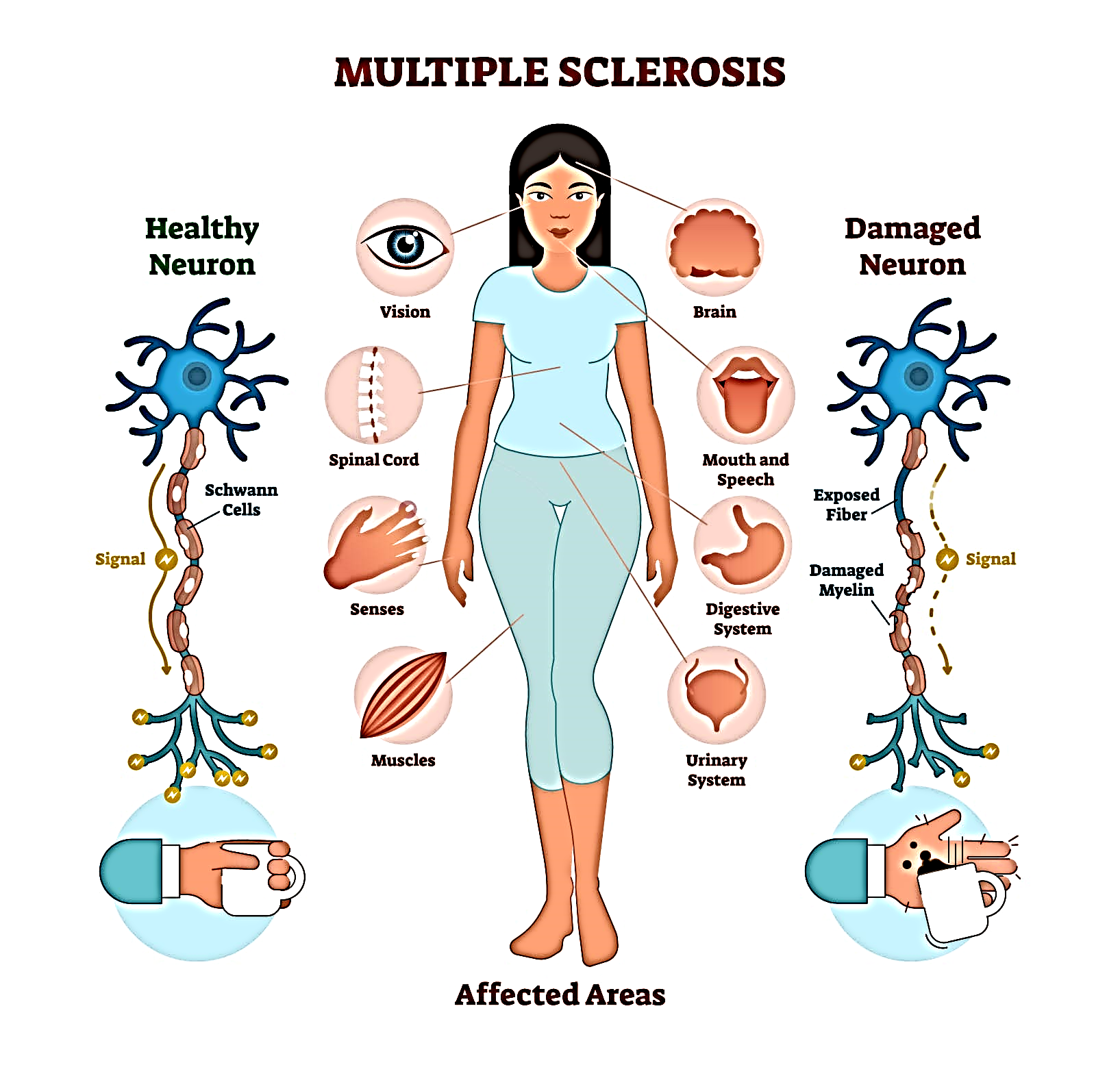
Multiple sclerosis is a disease that causes vision problems, numbness and tingling, muscle weakness, and other problems. It happens when the body’s infection-fighting system attacks and damages nerve cells and their connections in the brain and spinal cord. When the body’s infection-fighting system, called the “immune system,” attacks the body’s own cells, it is called an “autoimmune response.” It causes damage to myelin, the protective coating around the nerves. When myelin is damaged, messages can no longer be clearly transmitted from the brain and spinal cord to other parts of the body. Many people refer to multiple sclerosis as “MS.” INCIDENCE

  • Onset occurs between 20-40 years of age.
  • Women are more affected than men. (AANN, 2011).
  • Whites are more affected than Hispanics, blacks, or Asians.
  • Most prevalent in colder climates of North America & Europe.
  • Migration.


ETIOLOGY & RISK FACTORS

  • Exact cause is not known yet.
  • Most theories suggest that MS is an immunogenetic viral disease (with Epstein Barr virus).

Risk factors are:

  • Age (most of the time between 20-40 yrs).
  • Sex (women have more chance).
  • Family history (genetic susceptibility).
  • Certain infections (like Epsteinbarr virus).
  • Climate (more in cold climate areas).
  • Certain auto-immune diseases (higher risks with thyroid disease, type-1 DM or IBD).
  • Smoking.
  • Stress, fatigue.
  • Physical injury.
  • Pregnancy (may relating to stress to labour, or puerperium).


PATHOPHYSIOLOGY Due to etiological factors Activated T-cells (which recognise self Ag) expressed in CNS, & Macrophages (B-cells) enters the brain from peripherral circulation Production of inflammatory cytokines & reactive O2 species Inflammation Then activated T-cells & B-cells cause demyelination and destruction of oligodendrocytes Formation of plaque Causes scarring & destruction of sheath Compensatory system starts causing subsidation of edema & inflammation After that some remyelination process occurs which is often incomplete Multiple sclerosis. CLINICAL MANIFESTATIONS The course of illness varies from person to person.

  1. Fatigue is the lack of physical and mental energy that impacts daily tasks. Fatigue can be physical or mental and is not correlated to how much rest or sleep a person gets. It is one of the most common symptoms and impacts about 80 percent of people living with MS. It can be http://nursingfile.com/wp-content/uploads/2013/12/multiple-sclerosis-249x300.jpg the most debilitating factor, even for those who have minimal physical restrictions, and is one of the leading causes for people leaving the workforce.
  2. Heat intolerance in MS is a temporary worsening of symptoms with elevated body temperatures including hot and humid weather, exercising, sunbathing, or fevers. A small rise in body temperature (a quarter to a half a degree) can cause increased fatigue, tingling, blurry vision, or even the inability to walk. Most people living with MS have to avoid outdoor activity and/or use cooling garments to complete simple, daily activities due to this intolerance.
  3. Cognitive dysfunction affects high-level brain functions such as memory, attention/concentration, the ability to solve daily problems, understand and use language, and process information from different senses. Impaired cognition affects 50-65 percent of those living with MS and is another major reason for leaving the workforce early.
  4. Pain/abnormal sensation is a common symptom with MS and can be directly related to neuropathic pain (the disease process itself) or from musculoskeletal pain (changes to the body and immobility). The pain experience is unique to each person and can greatly limit his or her ability to participate in and enjoy socialization and activities. Those living with MS can also experience various abnormal sensations such as numbness and tingling, prickling, sharp/stabbing pains, hot/cold sensations, and burning pains which can also impact movement and daily function.
  5. Depression comes in various forms and can be one of the most common symptoms in MS, more common in people with MS than the general population. Depression can happen to anyone at any time during the disease course and does not correlate to disease severity, however it can greatly impact someone’s quality of life and ability to participate in daily activities.


COMPLICATIONS People with multiple sclerosis may also develop:

  • Muscle stiffness or spasms
  • Paralysis, typically in the legs
  • Problems with bladder, bowel or sexual function
  • Mental changes, such as forgetfulness or mood swings
  • Depression
  • Epilepsy

DIAGNOSTIC EVALUATION

  • There is no definitive test for MS.
  • Detailed history of episodes of neurologic dysfunction
  • Physical examination.

Other tests include:-

  • CSF evaluation (for presence of IgG antibody or oligoclonal bonding)
  • Evoked potentials of optic pathways & auditory system to assess presence of slowed nerve conduction.
  • MRI of brain and spinal cord (to determine the presence of MS plaques)
  • CT scan (to detect areas of demyelination, but with less detail as by MRI).


MEDICAL MANAGEMENT

• No exact cure.
• Aim is to prevent or postpone the long term disability (often evolves slowly over many years).
• The treatment falls into 3 categories:-
1. Treatment of acute relapses.
2. Treatment aimed at disease management.
3. Symptomatic treatment.

1. Treatment of acute relapse:-

  • Corticosteroid therapy ( anti-inflammatory & immunosuppressive property )
  • For example:
  • Methyl-prednisolone , (given I.V. or orally)
  • Azathioprine & cyclophosphamide (in severe cases)

2. Treat exacerbations:- (treatment aimed at disease management)

  • Interferon-Beta 1b
    • Betaseron, given subcutaneously. (antiviral & immuno-regulatory) (For ambulatory clients with relapsing –remitting).
  • Interferon Beta 1a
    • Avonex, (for treating replasing form of MS).
  • Glatiramer acetate
    • Copaxane, (for relapsing re-emitting MS).

3. Symptomatic treatment:-

  • For bladder dysfunction:
    • oxybutynin, propantheline.
  • For constipation:
    • psyllium hydrophilic mucilloid, suppositories.
  • For fatigue:
    • amantadine, modafinil .
  • For spasticity:
    • baclofen, diazefen, dantrolone.
  • For Tremor :
    • propanolol, phenobarbital, clonazepam.
  • For dysesthesias & trigeminal neurolgia:
    • carbamazepine, phenytoin, amitriptyline.
  • For dysesthesias:
    • Transcutaneous electrical nerve stimulation (TENS) is also helpful.

4. Nutritional therapy:-

  • Megavitamin therapy (cobalamin/vit. B12 and vit. C)
  • Low fat diet.
  • high roughage diet (to relieve constipation)

5. Other therapies:- (to improve neurological functioning)

  • Physical and speech therapies.
  • Exercise.
  • Water exercise.


SURGICAL MANAGEMENT

  • Deep brain stimulation:-
    • if other options have failed then a device is implanted that stimulates an area of brain. (in case of severe tremor in limbs).
  • Implantation of a drug catheter or pump:
    • a catheter is placed in lower spinal area to deliver a constant flow of drug like baclofen. (in case of severe pain or spasticity).

NURSING DIAGNOSIS:

    • Impaired physical mobility related to fatigue & weakness
    • Activity intolerance r/t weakness, dizziness, and unsteady gait
    • Self-esteem disturbance r/t loss of health & lifestyle changes


NURSING MANAGEMENT

    1. Promotes physical mobility – activity and rest
  • no vigorous physical exercise
  • frequent rest periods
  • walking and gait exercises
  • minimize spasticity and contractures – warm packs, daily muscle stretching
  • activities: swimming, stationary bike, progressive wt bearing
  • Minimize effects of immobility; skin integrity; cough and deep breathing exercises.
    1. Prevent injury – walk with feet wide apart, environment awareness and modification, gait training. Use of assistive devices – walker, cane etc.
    2. Promote bladder & bowel control – Urinal/bedpan readily available, po fluids intake schedule/voiding schedule, increase fiber in diet, intermittent self-catheterization
    3. Improve sensory and cognitive function:
  • Vision – eye patch for diplopia; prism glasses for reading; talking books
  • Speech – slurred, low volume, problems with phonation – speech therapist
  • Cognitive & emotional responses – forgetfulness, easily distracted, emotionally labile, social activities; hobbies.
    1. Development of coping strengths – education about diseases process; stress relief; network of services – social, speech, PT, psychological, homemaker/meal on wheels
    2. Improve self-care – assistive devices, raised toilet seat, shower bench, reached tongs, decrease physical and emotional stress, decrease exposure of extreme temperatures
    3. Adapting to sexual dysfunction – counseling, plan sexual activity, willingness to experiment.

Bronchiectasis

Bronchiectasis is a chronic, irreversible dilation of the bronchi and bronchioles. Under the new definition of COPD, it is considered a disease process separate from COPD (GOLD, 2008).
Bronchiectasis is a condition where the bronchial tubes of your lungs are permanently damaged, widened, and thickened.

Pathophysiology

Bronchiectasis is an obstructive lung condition in which there is destruction and widening of the large airways and abnormal bronchial wall thickening as a result of a recurring cycle of infection and inflammation (Ten Hacken 2010). Bronchiectasis is usually localised to one lung segment or lobe but may spread over time to other parts of the same lung as a result of unresolved infections. Exacerbations are associated with infections.
A number of conditions lead to bronchiectasis, including structural lung conditions, CF and other conditions associated with abnormal mucocillary clearance; retained inhaled foreign objects, tumours and obstructive lung conditions including COPD; an abnormal immune response; infections including tuberculosis, pneumonia, measles and whooping cough; and inflammatory bowel disease.

Clinical manifestations

  • Difficult breathing (dyspnea) due to the mucous production and irritation within the airways.
  • Productive, foul-smelling odorous cough, due to thick, difficult-to-expel, tenacious mucous, often with bacterial colonization.
  • Cough may be worse when lying down.
  • Recurrent bronchial infections.
  • Hemoptysis (blood-tinged or bloody mucous).
  • Loss of weight because patients are not eating well, due to respiratory changes and foul-smelling mucous with cough. Increased respiratory effort requires more calories to meet normal requirements.
  • Crackles or rhonchi on inspiration due to mucous build-up.
  • Anemia of chronic disease.
  • Cyanosis.
  • Clubbing of the fingers.

Causes
Bronchiectasis may be caused by a variety of conditions including:

  • Airway obstruction: Obstructions in the bronchi distend the wall permanently and impair mucociliary action.
  • Pulmonary infection: Pulmonary infection and obstruction of the bronchus or complications of long-term pulmonary infections cause bronchiectasis.
  • Genetic disorders: Genetic disorders such as cystic fibrosis causes the sputum to thicken in consistency and would ultimately obstruct the bronchi.
  • Idiopathic causes: There are causes that are unknown to medicine that cause bronchiectasis.

Complications
Potential complications include:

  • Atelectasi: Collapse of the alveoli is a common complication.
  • Pneumonia: Infection is recurrent in patients with bronchiectasis.
  • Empyema: Overproduction of sputum causes the bronchi to be filled with pus.

Diagnosis

  • Culture and sensitivity of sputum to identify bacteria and appropriate antibiotics.
  • Shadows in affected area of the lungs on the chest x-ray.
  • CT scan or high-resolution CT will show areas of bronchiectasis.
  • Decreased lung vital capacity on pulmonary function test.

TREATMENT
Treatment is focused on getting enough oxygen to meet current needs of the patient, expel mucous, and treat infections.

  • Supplemental oxygen to help meet body’s needs.
  • Postural drainage to assist with drainage of secretions.
  • Chest PT to loosen secretions.
  • Remove excessive secretions during a bronchoscopy.
  • Administer bronchodilators to help keep airways open:
    • albuterol, levalbuterol
  • Administer antibiotics to treat infection:
    • selected based on the results of a culture and sensitivity study

NURSING DIAGNOSES

  • Ineffective airway clearance
  • Imbalanced nutrition: less than what the body requires
  • Impaired gas exchange

NURSING INTERVENTION

  • Monitor respiratory rate, effort, breath sounds, skin color, and use of accessory muscles.
  • Perform chest percussion to help loosen secretions.
  • Explain to the patient:
    • That family member can perform chest PT.
    • How to do postural drainage.
    • How to administer oxygen.
    • How to properly administer medications.

See videos:

Bronchitis  Chronic Bronchitis

Abnormal breath sounds

Breath sounds are an important part of respiratory assessment and are usually assessed by the respiratory team.

Sound Characteristic Signs of
Wheezing Whistling sound, generally heard on expiration Asthma and airway obstruction
Stridor Snoring sound heard on inspiration Typical of obstruction, sputum plug or foreign body, anaphylactic reaction
Crackles A crackling or popping sound Collapsed alveoli popping open on inspiration
Rhonchi Snoring or rattling sounds Fluid partly blocking the bronchi; generally heard on expiration
Pleural friction A grating or rubbing sound heard on inspiration and expiration Indicative of pleural inflammation

General Respiratory Assessment


Focus Observe Normal adult values Abnormalities Indications
Respiration Quality, rate Rate: 12–18 per
minute
Bradypnoea <10
breaths per minute
Head injury
Narcotic overdose
Tachypnoea Following exertion
Pneumonia
Diabetic ketoacidosis
Pyrexia – rate of
breathing increases by
7 breaths per minute
for every 10°C rise in
the patient’s
temperature
Apnoea Respiratory arrest
Depth Shallow or deep Hyperventilation
Hypoventilation
(hypercapnia)
Rhythm Regular rhythm Cluster breathing –
normal breathing
interspersed with
apnoeic pauses
Neurological disorders
Cheyne–Stokes –
the rate changes
with increasing
periods of apnoea
End of life
Effort Unable to complete a sentence
Use of accessory
muscles
Muscle retraction –intercostal and
suprasternal muscles
Nasal flaring
Pneumonia
Airway inflammation/
obstruction
Emphysema
Trauma
Colour Skin colour –
check the
skin, nail beds, lips, tongue, ears and nose
Cyanosis – central or peripheral Central – hypoxaemia
Peripheral – perfusion
abnormality
Chest Shape Barrel chest Emphysema
Pigeon chest Rickets
Movement Symmetrical Asymmetrical Pneumothorax – air
trapped in the pleura
Extreme atelectasis

Chronic Obstructive Pulmonary Disease (COPD)

Key facts

  • Chronic obstructive pulmonary disease (COPD) is a progressive life-­threatening lung disease that causes breathlessness (initially with exertion) and predisposes to exacerbations and serious illness.
  • The Global Burden of Disease Study reports a prevalence of 251 million cases of COPD globally in 2016.
  • Globally, it is estimated that 3.17 million deaths were caused by the disease in 2015 (that is, 5% of all deaths globally in that year).
  • More than 90% of COPD deaths occur in low­ and middle-­income countries.
  • The primary cause of COPD is exposure to tobacco smoke (either active smoking or second­hand smoke).
  • Other risk factors include exposure to indoor and outdoor air pollution and occupational dusts and fumes.
  • Exposure to indoor air pollution can affect the unborn child and represent a risk factor for developing COPD later in life.
  • Some cases of COPD are due to long-term asthma.
  • COPD is likely to increase in coming years due to higher smoking prevalence and aging populations in many countries.
  • Many cases of COPD are preventable by avoidance or early cessation of smoking. Hence, it is important that countries adopt the WHO Framework Convention on Tobacco Control (WHO-FCTC) and implement the MPOWER package of measures so that non-smoking becomes the norm globally.
  • COPD is not curable, but treatment can relieve symptoms, improve quality of life and reduce the risk of death.

Chronic obstructive pulmonary disease (COPD) is a disease state characterized by airflow limitation that is not fully reversible. This newest definition of COPD, provided by the Global Initiative for Chronic Obstructive Lung Disease, provides a broad description that better explains this disorder and its signs and symptoms (National Institutes of Health [NIH], 2001).

Chronic obstructive pulmonary disease (COPD)

While previous definitions have included emphysema and chronic bronchitis under the umbrella classification of COPD, this was often confusing because most patients with COPD present with overlapping signs and symptoms of these two distinct disease processes.

 COPD may include diseases that cause airflow obstruction (eg, emphysema, chronic bronchitis) or a combination of these disorders. Other diseases such as cystic fibrosis, bronchiectasis, and asthma were previously classified as types of chronic obstructive lung disease. However, asthma is now considered a separate disorder and is classified as an abnormal airway condition characterized primarily by reversible inflammation. COPD can coexist with asthma. Both of these diseases have the same major symptoms; however, symptoms are generally more variable in asthma than in COPD.

People with COPD commonly become symptomatic during the middle adult years, and the incidence of COPD increases with age. Although certain aspects of lung function normally decrease with age (eg, vital capacity and forced expiratory volume in 1 second [FEV1]), COPD accentuates and accelerates these physiologic changes.

Pathophysiology

In COPD, the airflow limitation is both progressive and associated with an abnormal inflammatory response of the lungs to noxious particles or gases. The inflammatory response occurs throughout the airways, parenchyma, and pulmonary vasculature (NIH, 2001). Because of the chronic inflammation and the body’s attempts to repair it, narrowing occurs in the small peripheral airways. Over time, this injury-and-repair process causes scar tissue formation and narrowing of the airway lumen. Airflow obstruction may also be due to parenchymal destruction as seen with emphysema, a disease of the alveoli or gas exchange units.

In addition to inflammation, processes relating to imbalances of proteinases and antiproteinases in the lung may be responsible for airflow limitation. When activated by chronic inflammation, proteinases and other substances may be released, damaging the parenchyma of the lung. The parenchymal changes may also be consequences of inflammation, environmental, or genetic factors (eg, alpha1 antitrypsin deficiency).

Early in the course of COPD, the inflammatory response causes pulmonary vasculature changes that are characterized by thickening of the vessel wall. These changes may occur as a result of exposure to cigarette smoke or use of tobacco products or as a result of the release of inflammatory mediators (NIH, 2001).

Chronic Bronchitis

Chronic bronchitis, a disease of the airways, is defined as the presence of cough and sputum production for at least 3 months in each of 2 consecutive years. In many cases, smoke or other environmental pollutants irritate the airways, resulting in hypersecretion of mucus and inflammation. This constant irritation causes the mucus-secreting glands and goblet cells to increase in number, ciliary function is reduced, and more mucus is produced. The bronchial walls become thickened, the bronchial lumen is narrowed, and mucus may plug the airway. Alveoli adjacent to the bronchioles may become damaged and fibrosed, resulting in altered function of the alveolar macrophages. This is significant because the macrophages play an important role in destroying foreign particles, including bacteria. As a result, the patient becomes more susceptible to respiratory infection. A wide range of viral, bacterial, and mycoplasmal infections can produce acute episodes of bronchitis. Exacerbations of chronic bronchitis are most likely to occur during the winter.

Emphysema

In emphysema, impaired gas exchange (oxygen, carbon dioxide) results from destruction of the walls of overdistended alveoli. “Emphysema” is a pathological term that describes an abnormal distention of the air spaces beyond the terminal bronchioles, with destruction of the walls of the alveoli. It is the end stage of a process that has progressed slowly for many years. As the walls of the alveoli are destroyed (a process accelerated by recurrent infections), the alveolar surface area in direct contact with the pulmonary capillaries continually decreases, causing an increase in dead space (lung area where no gas exchange can occur) and impaired oxygen diffusion, which leads to hypoxemia. In the later stages of the disease, carbon dioxide elimination is impaired, resulting in increased carbon dioxide tension in arterial blood (hypercapnia) and causing respiratory acidosis. As the alveolar walls continue to break down, the pulmonary capillary bed is reduced.

Consequently, pulmonary blood flow is increased, forcing the right ventricle to maintain a higher blood pressure in the pulmonary artery. Hypoxemia may further increase pulmonary artery pressure. Thus, right-sided heart failure (cor pulmonale) is one of the complications of emphysema. Congestion, dependent edema, distended neck veins, or pain in the region of the liver suggests the development of cardiac failure.

Risk Factors for COPD

  • Exposure to tobacco smoke accounts for an estimated 80% to 90% of COPD cases (Rennard, 1998)
  • Passive smoking
  • Occupational exposure
  • Ambient air pollution
  • Genetic abnormalities, including a deficiency of alpha1-antitrypsin, an enzyme inhibitor that normally counteracts the destruction of lung tissue by certain other enzymes

Clinical Manifestations

COPD is characterized by three primary symptoms:

Katie Hrubec Hrubec Hrubec Burroughs @Megan Ward Ward Ward Anderson i guess i finally know what COPD is | Nursing school survival, Nursing mnemonics, Nurse

  • cough,
  • sputum production, and
  • dyspnea on exertion. These symptoms often worsen over time.
  • Chronic cough and sputum production often precede the development of airflow limitation by many years. However, not all individuals with cough and sputum production will develop COPD.
  • Dyspnea may be severe and often interferes with the patient’s activities.
  • Weight loss is common because dyspnea interferes with eating, and the work of breathing is energy-depleting.

Often the patient cannot participate in even mild exercise because of dyspnea; as COPD progresses, dyspnea occurs even at rest. As the work of breathing increases over time, the accessory muscles are recruited in an effort to breathe. The patient with COPD is at risk for respiratory insufficiency and respiratory infections, which in turn increase the risk for acute and chronic respiratory failure.

  • In COPD patients with a primary emphysematous component, chronic hyperinflation leads to the “barrel chest” thorax configuration. This results from fixation of the ribs in the inspiratory position (due to hyperinflation) and from loss of lung elasticity. Retraction of the supraclavicular fossae occurs on inspiration, causing the shoulders to heave upward. In advanced emphysema, the abdominal muscles also contract on inspiration.

Complications

There are two major life-threatening complications of COPD: respiratory insufficiency and failure.

  • Respiratory failure. The acuity and the onset of respiratory failure depend on baseline pulmonary function, pulse oximetry or arterial blood gas values, comorbid conditions, and the severity of other complications of COPD.
  • Respiratory insufficiency. This can be acute or chronic, and may necessitate ventilator support until other acute complications can be treated.

Assessment and Diagnostic Findings

Diagnosis and assessment of COPD must be done carefully since the three main symptoms are common among chronic pulmonary disorders.

  • Health history. The nurse should obtain a thorough health history from patients with known or potential COPD.
  • Pulmonary function studies. Pulmonary function studies are used to help confirm the diagnosis of COPD, determine disease severity, and monitor disease progression.
  • Spirometry. Spirometry is used to evaluate airway obstruction, which is determined by the ratio of FEV1 to forced vital capacity.
  • ABG. Arterial blood gas measurement is used to assess baseline oxygenation and gas exchange and is especially important in advanced COPD.
  • Chest x-ray. A chest x-ray may be obtained to exclude alternative diagnoses.
  • CT scan. Computed tomography chest scan may help in the differential diagnosis.
  • Screening for alpha1-antitrypsin deficiency. Screening can be performed for patients younger than 45 years old and for those with a strong family history of COPD.
  • Chest x-ray: May reveal hyperinflation of lungs, flattened diaphragm, increased retrosternal air space, decreased vascular markings/bullae (emphysema), increased bronchovascular markings (bronchitis), normal findings during periods of remission (asthma).
  • Pulmonary function tests: Done to determine cause of dyspnea, whether functional abnormality is obstructive or restrictive, to estimate degree of dysfunction and to evaluate effects of therapy, e.g., bronchodilators. Exercise pulmonary function studies may also be done to evaluate activity tolerance in those with known pulmonary impairment/progression of disease.
  • The forced expiratory volume over 1 second (FEV1): Reduced FEV1 not only is the standard way of assessing the clinical course and degree of reversibility in response to therapy, but also is an important predictor of prognosis.
  • Total lung capacity (TLC), functional residual capacity (FRC), and residual volume (RV): May be increased, indicating air-trapping. In obstructive lung disease, the RV will make up the greater portion of the TLC.
  • Arterial blood gases (ABGs): Determines degree and severity of disease process, e.g., most often Pao2is decreased, and Paco2 is normal or increased in chronic bronchitis and emphysema, but is often decreased in asthma; pH normal or acidotic, mild respiratory alkalosis secondary to hyperventilation (moderate emphysema or asthma).
  • DL CO test: Assesses diffusion in lungs. Carbon monoxide is used to measure gas diffusion across the alveocapillary membrane. Because carbon monoxide combines with hemoglobin 200 times more easily than oxygen, it easily affects the alveoli and small airways where gas exchange occurs. Emphysema is the only obstructive disease that causes diffusion dysfunction.
  • Bronchogram: Can show cylindrical dilation of bronchi on inspiration; bronchial collapse on forced expiration (emphysema); enlarged mucous ducts (bronchitis).
  • Lung scan: Perfusion/ventilation studies may be done to differentiate between the various pulmonary diseases. COPD is characterized by a mismatch of perfusion and ventilation (i.e., areas of abnormal ventilation in area of perfusion defect).
  • Complete blood count (CBC) and differential: Increased hemoglobin (advanced emphysema), increased eosinophils (asthma).
  • Blood chemistry: alpha1-antitrypsin is measured to verify deficiency and diagnosis of primary emphysema.
  • Sputum culture: Determines presence of infection, identifies pathogen.
  • Cytologic examination: Rules out underlying malignancy or allergic disorder.
  • Electrocardiogram (ECG): Right axis deviation, peaked P waves (severe asthma); atrial dysrhythmias (bronchitis), tall, peaked P waves in leads II, III, AVF (bronchitis, emphysema); vertical QRS axis (emphysema).
  • Exercise ECG, stress test: Helps in assessing degree of pulmonary dysfunction, evaluating effectiveness of bronchodilator therapy, planning/evaluating exercise program.

Medical Management

Healthcare providers perform medical management by considering the assessment data first and matching the appropriate intervention to the existing manifestation.

Pharmacologic Therapy

  • Bronchodilators. Bronchodilators relieve bronchospasm by altering the smooth muscle tone and reduce airway obstruction by allowing increased oxygen distribution throughout the lungs and improving alveolar ventilation.
  • Corticosteroids. A short trial course of oral corticosteroids may be prescribed for patients to determine whether pulmonary function improves and symptoms decrease.
  • Other medications. Other pharmacologic treatments that may be used in COPD include alpha1-antitrypsin augmentation therapy, antibiotic agents, mucolytic agents, antitussive agents, vasodilators, and narcotics.

Management of Exacerbations

  • Optimization of bronchodilator medications is first-line therapy and involves identifying the best medications or combinations of medications taken on a regular schedule for a specific patient.
  • Hospitalization. Indications for hospitalization for acute exacerbation of COPD include severe dyspnea that does not respond to initial therapy, confusion or lethargy, respiratory muscle fatigue, paradoxical chest wall movement, and peripheral edema.
  • Oxygen therapy. Upon arrival of the patient in the emergency room, supplemental oxygen therapy is administered and rapid assessment is performed to determine if the exacerbation is life-threatening.
  • Antibiotics. Antibiotics have been shown to be of some benefit to patients with increased dyspnea, increased sputum production, and increased sputum purulence.

Surgical Management

Patients with COPD also have options for surgery to improve their condition.

  • Bullectomy. Bullectomy is a surgical option for select patients with bullous emphysema and can help reduce dyspnea and improve lung function.
  • Lung Volume Reduction Surgery. Lung volume reduction surgery is a palliative surgery in patients with homogenous disease or disease that is focused in one area and not widespread throughout the lungs.
  • Lung Transplantation. Lung transplantation is a viable option for definitive surgical treatment of end-stage emphysema.

Nursing Management

Management of patients with COPD should be incorporated with teaching and improving the respiratory status of the patient.

Nursing Assessment

Assessment of the respiratory system should be done rapidly yet accurately.

  • Assess patient’s exposure to risk factors.
  • Assess the patient’s past and present medical history.
  • Assess the signs and symptoms of COPD and their severity.
  • Assess the patient’s knowledge of the disease.
  • Assess the patient’s vital signs.
  • Assess breath sounds and pattern.

Diagnosis

Diagnosis of COPD would mainly depend on the assessment data gathered by the healthcare team members.

  • Impaired gas exchange due to chronic inhalation of toxins.
  • Ineffective airway clearance related to bronchoconstriction, increased mucus production, ineffective cough, and other complications.
  • Ineffective breathing pattern related to shortness of breath, mucus, bronchoconstriction, and airway irritants.
  • Self-care deficit related to fatigue.
  • Activity intolerance related to hypoxemia and ineffective breathing patterns.

Planning & Goals

Goals to achieve in patients with COPD include:

  • Improvement in gas exchange.
  • Achievement of airway clearance.
  • Improvement in breathing pattern.
  • Independence in self-care activities.
  • Improvement in activity intolerance.
  • Ventilation/oxygenation adequate to meet self-care needs.
  • Nutritional intake meeting caloric needs.
  • Infection treated/prevented.
  • Disease process/prognosis and therapeutic regimen understood.
  • Plan in place to meet needs after discharge.

Nursing Priorities

  1. Maintain airway patency.
  2. Assist with measures to facilitate gas exchange.
  3. Enhance nutritional intake.
  4. Prevent complications, slow progression of condition.
  5. Provide information about disease process/prognosis and treatment regimen.

Nursing Interventions

Patient and family teaching is an important nursing intervention to enhance self-management in patients with any chronic pulmonary disorder.

To achieve airway clearance:

  • The nurse must appropriately administer bronchodilators and corticosteroids and become alert for potential side effects.
  • Direct or controlled coughing. The nurse instructs the patient in direct or controlled coughing, which is more effective and reduces fatigue associated with undirected forceful coughing.

To improve breathing pattern:

  • Inspiratory muscle training. This may help improve the breathing pattern.
  • Diaphragmatic breathing. Diaphragmatic breathing reduces respiratory rate, increases alveolar ventilation, and sometimes helps expel as much air as possible during expiration.
  • Pursed lip breathing. Pursed lip breathing helps slow expiration, prevents collapse of small airways, and control the rate and depth of respiration.

To improve activity intolerance:

  • Manage daily activities. Daily activities must be paced throughout the day and support devices can be also used to decrease energy expenditure.
  • Exercise training. Exercise training can help strengthen muscles of the upper and lower extremities and improve exercise tolerance and endurance.
  • Walking aids. Use of walking aids may be recommended to improve activity levels and ambulation.

To monitor and manage potential complications:

  • Monitor cognitive changes. The nurse should monitor for cognitive changes such as personality and behavior changes and memory impairment.
  • Monitor pulse oximetry values. Pulse oximetry values are used to assess the patient’s need for oxygen and administer supplemental oxygen as prescribed.
  • Prevent infection. The nurse should encourage the patient to be immunized against influenza and S. pneumonia because the patient is prone to respiratory infection.

Evaluation

During evaluation, the effectiveness of the care plan would be measured if goals were achieved in the end and the patient:

  • Identifies the hazards of cigarette smoking.
  • Identifies resources for smoking cessation.
  • Enrolls in smoking cessation program.
  • Minimizes or eliminates exposures.
  • Verbalizes the need for fluids.
  • Is free of infection.
  • Practices breathing techniques.
  • Performs activities with less shortness of breath.

Discharge and Home Care Guidelines

It is important for the nurse to assess the knowledge of patient and family members about self-care and the therapeutic regimen.

  • Setting goals. If the COPD is mild, the objectives of the treatment are to increase exercise tolerance and prevent further loss of pulmonary function, while if COPD is severe, these objectives are to preserve current pulmonary function and relieve symptoms as much as possible.
  • Temperature control. The nurse should instruct the patient to avoid extremes of heat and cold because heat increases the temperature and thereby raising oxygen requirements and high altitudes increase hypoxemia.
  • Activity moderation. The patient should adapt a lifestyle of moderate activity and should avoid emotional disturbances and stressful situations that might trigger a coughing episode.
  • Breathing retraining. The home care nurse must provide the education and breathing retraining necessary to optimize the patient’s functional status.

Documentation Guidelines

Documentation is an essential part of the patient’s chart because the interventions and medications given and done are reflected on this part.

  • Document assessment findings including respiratory rate, character of breath sounds; frequency, amount and appearance of secretions laboratory findings and mentation level.
  • Document conditions that interfere with oxygen supply.
  • Document plan of care and specific interventions.
  • Document liters of supplemental oxygen.
  • Document client’s responses to treatment, teaching, and actions performed.
  • Document teaching plan.
  • Document modifications to plan of care.
  • Document attainment or progress towards goals.