Anatomy and Physiology MCQs/BCQs

1. Bone and cartilage is a type of:
A. Nervous tissue
B. Muscular tissue
C. Epithelial tissue
D. Endocrine tissue
E. Connective tissue

2. Color of the skin, due to the presence of:
A. Collagen
B. Langerhans cells
C. Melanocytes
D. Merkel cells
E. Keratinocytes

3. Anosmia is loss of sense of
A. vision
B. hearing
C. smell
D. taste

4. The maximum volume air which can be moved into and out of the lungs is known as:
A. Total lung capacity
B. Inspiratory capacity
C. Vital capacity
D. Functional residual capacity

5. Regarding eye, sensory receptors for vision are:
A. Ciliary body
B. Rods and cones
C. Olfactory cells
D. Lens

6. The basic structure and functional unit of nervous system is:
A. Schwann Cells
B. Neurons
C. Astrocytes
D. Microglia

7. All of the following are the functions of oxytocin; except
A. Ejection of milk
B. Parturition
C. Fertilization
D. Formation of milk

8. A student identifying histological section under microscope. The tissue was multilayered. The upper most layer is squamous in shape. What type of epithelium it is?
A. Simple squamous epithelium
B. Transitional epithelium
C. Stratified squamous epithelium
D. Stratified cuboidal epithelium
E. Pseudostratified epithelium

9. Common iliac artery supplies the:
A. Lower limb
B. Abdomen
C. Thorax
D. Upper limb
E. Head and neck

10. Which of the following bone forms the axial skeleton?
A. Humerus
B. Radius
C. Femur
D. Ulna
E. Sternum

11. Center of micturition reflex is located in:
A. Lumber segment of cord
B. Cerebral cortex
C. Brainstem (pontine micturition center)
D. Sacral segment of spinal cord

A girl moves the upper limb in all directions during exercise, what type of movement she performed?
A. Abduction
B. Circumduction
C. Flexion
D. Adduction
E. Extension

12. Adrenal medulla secretes:
A. Adrenaline and noradrenaline
B. mineralocorticoid
C. Glucocorticoid
D. Androgens

13. Short bones are present in which part of the human body?
A. Palm and sole
B. Leg
C. Upper arm
D. Thigh region
E. Forearm

14. The hormone which promotes tissue growth and regulates metabolisms:
A. Aldosterone
B. Thyroid hormone
C. Prolactin
D. Growth hormone

15. Which of the following chamber of the heart contain the sinoatrial node(SA)?
A. Left atrium
B. Left auricle
C. Right atrium
D. Right ventricle
E. Left ventricle

16. The cells form the myelin sheath in the central nervous system are:
A. Ependymal cells
B. Microglial cells
C. Astrocytes
D. Oligodendrocytes
E. Schwann cells

17. Which one of the following hormone is secreted by posterior pituitary gland?
A. Growth hormone
B. Antidiuretic hormone
C. Thyroid stimulating hormone
D. Follicle stimulating hormone

18. The most common synapse in CNS is:
A. Mechanical Synapse
B. Chemical Synapse
C. Gap Junctions
D. Electrical Synapse

19. Central nervous system is made up of:
A. Peripheral nerves
B. None of these
C. Brain and spinal cord
D. Somatic nerves

20. Exchange of gases by diffusion between blood and body cells is known as:
A. Alveolar ventilation
B. Internal respiration
C. Pulmonary ventilation
D. External respiration

21. How much percentage of oxygen is transported inform of oxyhemoglobin?
A. 1.5 %
B. 60 %
C. 98.5 %
D. 40 %

22. Dorsiflexion movement occur at which of the following joint:
A. Ankle joint
B. Shoulder joint
C. Knee joint
D. Elbow joint
E. Wrist joint

23. A body is divided into anterior and posterior half by which of the following imaginary plane
A. Midsagittal plane
B. Coronal plane
C. Right median plane
D. Left median plane
E. Para-median plane

24. Which of the following bones united by the sutures?
A. Sesamoid bones
B. Skull bones
C. Long bones
D. Tarsal bones
E. Carpal bones

25. Renin is secreted by:
A. Juxtaglomerular cells(JG)
B. PCT
C. DCT
D. Vasa recta

26. Main Muscle of quiet inspiration is:
A. Internal Intercostal
B. External Intercostal
C. Diaphragm
D. Abdominals

27. Surfactant is secreted by:
A. Type I Pneumocystis
B. Goblet Cells
C. Type IV Pneumocystis
D. Type II Pneumocystis

28. Shoulder joint is a type of:
A. Cartilaginous joint
B. Syndesmosis
C. Synovial joint
D. Fibrous joint
E. Secondary cartilaginous joint

29. The release of thyroid hormones (T3 andT4) in blood is stimulated by:
A. FSH
B. ACTH
C. TSH
D. LH

30. Skin is lined by:
A. Stratified squamous epithelium
B. Cuboidal epithelium
C. Transitional epithelium
D. Pseudostratified epithelium
E. Columnar epithelium

31. Pharynx continue with the esophagus at the level of:
A. 2nd thoracic vertebra
B. 6th cervical vertebra
C. 3rd cervical vertebra
D. 2nd cervical vertebra
E. 4th cervical vertebra

32. Somatic, cutaneous senses which originates from the skin are:
A. Chemoreceptors
B. Special senses
C. Pain, touch, cold and heat
D. proprioceptors

33. Heart receives the parasympathetic supply by means of:
A. Cranial nerves
B. Vagus nerve
C. Sympathetic plexus
D. Cervical nerves

34. A student was standing in class with folding both arms which one of the following movement she did performed?
A. Medial rotation at shoulder region
B. Flexion at shoulder region
C. Extension at shoulder region
D. Circumduction at shoulder region
E. Lateral rotation at shoulder region

35. Which of the following part is not included in large intestine?
A. Cecum
B. Descending colon
C. Duodenum
D. Ascending colon
E. Transverse colon

36. System of the body which is NOT necessary for survival is:
A. Central nervous system
B. Cardiovascular system
C. Reproductive system
D. Respiratory system

37. Al are the phases of menstrual cycle except:
A. Luteal phase
B. Secretory phase
C. Menstrual phase
D. Proliferative phase

38. Superior venacava is formed by the union of:
A. Cardiac veins
B. Common iliac veins
C. Brachiocephalic veins
D. Internal jugular veins
E. Azygous veins


Answer Key

1 E

2 C

3 C

4 C

5 B

6 B

7 A

8 C

9 A

10 B

11 C

12 A

13 A

14 A

15 D

16 E

17 C

18 B

19 C

20 B

21 C

22 A

23 A

24 B

25 B

26 B

27D

28 C

29 C

30 A

31 C

32 A

33 B

34 B

35 C

36 C

37 A

38 C


Features of sensory nerve and motor nerve:

Sensory nerves:

An afferent nerve conveying impulses that are processed by the central nervous system to become part of the organism’s perception of itself and of its environment.

The main function of the sensory nervous system is to inform the central nervous system about stimuli impinging on us from the outside or within us. By doing so, it informs us about any changes in the internal and external environment.

Sensory nerves contain only afferent fibers, long dendrites of sensory neurons. Motor nerves have only efferent fibers, long axons of motor neurons. Mixed nerves contain both types of fibers. A connective tissue sheath called the epineurium surrounds each nerve.

Motor nerves:

A motor nerve is a nerve located in the central nervous system (CNS), usually the spinal cord, that sends motor signals from the CNS to the muscles of the body. This is different from the motor neuron, which includes a cell body and branching of dendrites, while the nerve is made up of a bundle of axons.

Motor nerves have only efferent fibers, long axons of motor neurons.

Lateral wall of nose

Short essay on lateral wall of nose:

The lateral wall of the nasal cavity is a region of the nasopharynx essential for humidifying and filtering the air we breathe in nasally.

Here we can find a structure called agger nasi. The agger nasi is also referred to as the ‘nasoturbinal concha’ or ‘nasal ridge.’ It can be described as a small mound or ridge found in the lateral side of the nasal cavity. The structure is located midway along the anterior aspect of the middle nasal concha. An abnormally enlarged form may restrict the drainage of the frontal sinus by obstructing the frontal recess area.

The lateral cartilage (upper lateral cartilage, lateral process of septal nasal cartilage) is situated below the inferior margin of the nasal bone, and is flattened, and triangular in shape.

Surfactant

What is surfactant and what is its function?

Surfactant is a mixture of fat and proteins made in the lungs. Surfactant coats the alveoli (the air sacs in the lungs where oxygen enters the body). This prevents the alveoli from sticking together when your baby exhales (breathes out).

Its classically known role is to decrease surface tension in alveolar air spaces to a degree that facilitates adequate ventilation of the peripheral lung.

Physiology of the eye

What is the physiology of the eye?

The eye is composed of a series of lenses and spaces that give focus to images, just as a camera does. It is composed of the vitreous humor, aqueous humor, the crystalline lens, and the cornea, and each of these has its own refraction index (the average being 1.34, because of the content of these tissues).

Physiology of the Eye

The primary function of the eye is to form a clear image of objects in our environment. These images are transmitted to the brain through the optic nerve and the posterior visual pathways.

The various tissues of the eye and its adnexa are thus designed to facilitate this function.

The Eyelids

Functions include: (1) protection of the eye from mechanical trauma, extremes of temperature and bright light, and (2) maintenance of the normal precorneal tear film, which is important for maintenance of corneal health and clarity.

The Tear Film

The tear film consists of three layers: the mucoid, aqueous and oily layers.

The mucoid layer lies adjacent to the corneal epithelium.It improves the wetting properties of the tears.

The Cornea

The primary function of the cornea is refraction. In order to perform this function, the cornea requires the following:

  • transparency
  • smooth and regular surface
  • spherical curvature of proper refractive power
  • appropriate index of refraction.

The Aqueous Humour

The aqueous humour is an optically clear solution of electrolytes (in water) that fills the space between the cornea and the lens. Normal volume is 0.3 ml. Its function is to nourish the lens and cornea.

The Vitreous Body

The vitreous consists of a three-dimensional network of collagen fibers with the interspaces filled with polymerized hyaluronic acid molecules, which are capable of holding large quantities of water.

The Lens

The lens, like the cornea, is transparent. It is avascular and depends on the aqueous for nourishment.

It has a thick elastic capsule, which prevents molecules (e.g., proteins) moving into or out of it.

The Ciliary Body

The ciliary muscle (within the ciliary body) is a mass of smooth muscle, which runs circumferentially inside the globe and is attached to the scleral spur anteriorly.

Accommodation

Accommodation is the process whereby relaxation of zonular fibers allows the lens to become more globular, thereby increasing its refractive power.

The Retina

This is the “photographic film” of the eye that converts light into electrical energy (transduction) for transmission to the brain.

Laryngeal Cartilages

The larynx consists of nine laryngeal cartilages: three are single (epiglottic, thyroid, cricoid) and three are paired (arytenoid, corniculate, and cuneiform).

  • Thyroid:
    • Largest of the cartilages
    • It is composed of two plate-like laminae that fuse on the anterior side of the cartilage to form a peak, called the laryngeal prominence, known as the Adam’s apple.
    • Its posterior border is elongated both inferiorly and superiorly to form the superior horn of thyroid cartilage and inferior horn of thyroid cartilage.
  • Cricoid:
    • Only laryngeal cartilage to form a complete ring
  • Epiglottic cartilage:
    • Consists of elastic cartilage, giving flexibility to the epiglottis
    • Almost entirely covered in mucosa
    • Its stalk projects superiorly and attaches to the posterior aspect of the tongue, so that during swallowing the epiglottis will move to cover the respiratory opening, thus keeping food out of the lower respiratory tubules
  • Arytenoid:
    • Pyramid shaped
    • Anchor the vocal cords
  • Corniculate:
    • Attach to the apices of the arytenoid cartilages
  • Cuneiform:
    • Do not directly attach to other cartilages

Formation of Urine

The nephrons of the kidneys process blood and create urine through a process of filtration, reabsorption, and secretion. Urine is about 95% water and 5% waste products. Nitrogenous wastes excreted in urine include urea, creatinine, ammonia, and uric acid.

Urine formation depends on three functions:

  • Filtration is accomplished by the movement of fluids from the blood into the Bowman’s Capsule
  • Reabsorption involves the transfer of essential solutes and water from the nephron back into the blood
  • Secretion involves the movement of materials from the blood back into the nephron
  • For a detailed diagram and summary, see Fig. 1 on p. 350 and Table 2 on p. 351

Filtration

  • Blood running through the afferent arteriole into the glomerulus is under high pressure (65 mmHg compared to 25 mmHg normally found in capillary beds)
  • Most dissolved solutes (see Table 1, p. 349 for a list) pass through the walls of the glomerulus into the Bowman’s capsule

Reabsorption

  • On average, 600 mL of fluid flows through the kidneys every minute
  • About 20% (or 120 mL) is filtered into the nephron
  • If all of that fluid left in urine, dehydration would be a constant danger
  • Fortunately, only 1 mL of urine is formed for every 120 mL, meaning that 119 mL of fluids and solutes are reabsorbed
  • Selective reabsorption occurs by both active and passive transport
  • Carrier molecules move Na+ ions across the cell membranes of the cells that line the nephron
  • Negative ions (Cl- and HCO3-) follow the positive Na+ ions by charge attraction
  • Many mitochondria supply energy needed for active transport
  • Reabsorption occurs until the threshold level of a substance is reached
  • Excess (like NaCl) remains in the nephron and is excreted with urine
  • Other molecules are actively transported from the proximal tubule
  • Glucose and amino acids attach to specific carrier molecules, which shuttle them out of the nephron and into the blood
  • The amount of solute that can be reabsorbed is limited
  • Ex – individuals with high blood glucose will excrete some in their urine
  • The solutes that are actively transported out of the nephron create an osmotic gradient that draws water in from the nephron
  • A second osmotic force, created by the proteins not filtered into the nephron, also help reabsorption
  • The proteins remain in the blood stream and draw water from the interstitial fluid into the blood
  • As water is reabsorbed from the nephron, the remaining solutes become more concentrated
  • Molecules like urea and uric acid will diffuse from the nephron back into the blood

Secretion

  • Secretion is the movement of wastes from the blood into the nephron
  • Nitrogen containing wastes, excess H+ ions, and minerals like K+ ions are examples of substances secreted
  • Drugs (like penicillin) can also be secreted
  • Cells loaded with mitochondria line the distal tubule, providing energy for active transport

What is vital capacity and its importance?

What is vital capacity and its importance?

Vital capacity (VC) is the maximum amount of air a person can expel from the lungs after a maximum inspiration. It is equal to the sum of inspiratory reserve volume, tidal volume, and expiratory reserve volume i.e, VC = IRV + TV + ERV.

The breathing capacity of the lungs expressed as the number of cubic inches or cubic centimeters of air that can be forcibly exhaled after a full inspiration. It is about 3.5 – 4.5 liters in the human body.

It promotes the act of supplying fresh air and getting rid of foul air, thereby increasing the gaseous exchange between the tissues and the environment. Thus, the greater the VC, the more is the energy available to the body. VC of a person gives important clues for diagnosing a lung problem.

Its measurement helps the doctor to decide about the possible causes of the diseases and about the line of treatment.

It determines the stamina of sportsperson and mountain climbers. Sportsperson and mountain dwellers have a higher vital capacity. Young persons have more vital capacity than aged.

Homeostasis

The term homeostasis is used by physiologists to mean maintenance of nearly constant conditions in the internal environment. Essentially all organs and tissues of the body perform functions that help maintain these relatively constant conditions. For instance, the lungs provide oxygen to the extracellular fluid to replenish the oxygen used by the cells, the kidneys maintain constant ion concentrations, and the gastrointestinal system provides nutrients.

The term homeostasis comes from the Ancient Greek ὅμοιος (hómoios (homeo), meaning “similar”), from στημι (hístēmi (stasis), “standing still”) and stasis, from στάσις (stásis, meaning “standing”). The concept of homeostasis was first described in 1865 by Claude Bernard, a French physiologist. However, the term was coined later in 1962 by the American physiologist Walter Bradford Cannon.

Concept of Extracellular and Intracellular fluid

  • Intracellular fluid—fluid contained within all of the cells of the body
  • Extracellular fluid—fluid outside the cells of the body and is the intern
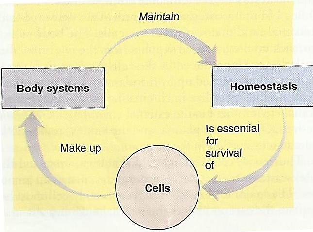
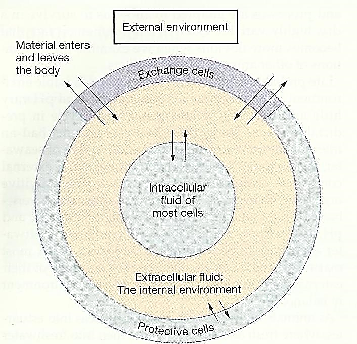
al environment in which the cells live. It is composed of plasma and interstitial fluid.


Interdependent relationship of cells, systems and homeostasis

Factors homeostatically regulated

  • Concentration of nutrient molecules
  • Concentration of CO2 andO2
  • Concentration of waste products
  • pH
  • Concentration of water, salt and other electrolytes
  • Temperature
  • Volume and pressure

Contribution of body systems to Homeostasis

  • The Circulatory System
    • Extracellular fluid is transported through all parts of the body in two stages. The first stage is movement of blood through the body in the blood vessels, and the second is movement of fluid between the blood capillaries and the intercellular spaces between the tissue cells.
    • All the blood in the circulation traverses the entire circulatory circuit an average of once each minute when the body is at rest and as many as six times each minute when a person is extremely active.
    • As blood passes through the blood capillaries, continual exchange of extracellular fluid also occurs between the plasma portion of the blood and the interstitial fluid that fills the intercellular spaces.
  • The Digestive System
    • A large portion of the blood pumped by the heart also passes through the walls of the gastrointestinal tract. Here different dissolved nutrients, including carbohydrates, fatty acids, and amino acids, are absorbed from the ingested food into the extracellular fluid of the blood.
    • Undigested material that enters the gastrointestinal tract and some waste products of metabolism are eliminated in the feces.
  • The Respiratory System
    • The blood picks up oxygen in the alveoli, thus acquiring the oxygen needed by the cells. The membrane between the alveoli and the lumen of the pulmonary capillaries, the alveolar membrane, is only 0.4 to 2.0 micrometers thick, and oxygen rapidly diffuses by molecular motion through this membrane into the blood.
    • Removal of Carbon Dioxide by the Lungs. At the same time that blood picks up oxygen in the lungs, carbon dioxide is released from the blood into the lung alveoli; the respiratory movement of air into and out of the lungs carries the carbon dioxide to the atmosphere. Carbon dioxide is the most abundant of all the end products of metabolism.
  • The Urinary System
    • Passage of the blood through the kidneys removes from the plasma most of the other substances besides carbon dioxide that are not needed by the cells.
    • These substances include different end products of cellular metabolism, such as urea and uric acid; they also include excesses of ions and water from the food that might have accumulated in the extracellular fluid.
    • The kidneys perform their function by first filtering large quantities of plasma through the glomeruli into the tubules and then reabsorbing into the blood those substances needed by the body, such as glucose, amino acids, appropriate amounts of water, and many of the ions. Most of the other substances that are not needed by the body, especially the metabolic end products such as urea, are reabsorbed poorly and pass through the renal tubules into the urine.
  • The Musculoskeletal system
    • How does the musculoskeletal system contribute to homeostasis? The answer is obvious and simple: Were it not for the muscles, the body could not move to the appropriate place at the appropriate time to obtain the foods required for nutrition. The musculoskeletal system also provides motility for protection against adverse surroundings, without which the entire body, along with its homeostatic mechanisms, could be destroyed instantaneously.
  • The Integumentary System
    • The skin and its various appendages, including the hair, nails, glands, and other structures, cover, cushion, and protect the deeper tissues and organs of the body and generally provide a boundary between the body’s internal environment and the outside world. The integumentary system is also important for temperature regulation and excretion of wastes and it provides a sensory interface between the body and the external environment. The skin generally comprises about 12 to 15 percent of body weight.
  • The Immune System
    • The immune system consists of the white blood cells, tissue cells derived from white blood cells, the thymus, lymph nodes, and lymph vessels that protect the body from pathogens such as bacteria, viruses, parasites, and fungi. The immune system provides a mechanism for the body to (1) distinguish its own cells from foreign cells and substances and (2) destroy the invader by phagocytosis or by producing sensitized lymphocytes or specialized proteins (e.g., antibodies) that either destroy or neutralize the invader.
  • The Nervous System
    • The nervous system is composed of three major parts: the sensory input portion, the central nervous system (or integrative portion), and the motor output portion. Sensory receptors detect the state of the body or the state of the surroundings. For instance, receptors in the skin apprise one whenever an object touches the skin at any point. The eyes are sensory organs that give one a visual image of the surrounding area. The ears are also sensory organs. The central nervous system is composed of the brain and spinal cord. The brain can store information, generate thoughts, create ambition, and determine reactions that the body performs in response to the sensations. Appropriate signals are then transmitted through the motor output portion of the nervous system to carry out one’s desires.
  • The Endocrine System
    • Located in the body are eight major endocrine glands that secrete chemical substances called hormones. Hormones are transported in the extracellular fluid to all parts of the body to help regulate cellular function. For instance, thyroid hormone increases the rates of most chemical reactions in all cells, thus helping to set the tempo of bodily activity. Insulin controls glucose metabolism; adrenocortical hormones control sodium ion, potassium ion, and protein metabolism; and parathyroid hormone controls bone calcium and phosphate. Thus, the hormones provide a system for regulation that complements the nervous system.
  • Reproductive system
    • Sometimes reproduction is not considered a homeostatic function. It does, however, help maintain homeostasis by generating new beings to take the place of those that are dying. This may sound like a permissive usage of the term homeostasis, but it illustrates that, in the final analysis, essentially all body structures are organized such that they help maintain the automaticity and continuity of life.

Homeostatic Control System

It is a functionally interconnected network of body components that operate to maintain a given physical or chemical factor in the internal environment relatively constant around an optimal level.

The human body has thousands of control systems. The most intricate of these are the genetic control systems that operate in all cells to help control intracellular function and extracellular functions.

Many other control systems operate within the organs to control functions of the individual parts of the organs; others operate throughout the entire body to control the interrelations between the organs. For instance, the respiratory system, operating in association with the nervous system, regulates the concentration of carbon dioxide in the extracellular fluid. The liver and pancreas regulate the concentration of glucose in the extracellular fluid, and the kidneys regulate concentrations of hydrogen, sodium, potassium, phosphate, and other ions in the extracellular fluid. Can be classified as:

  • Intrinsic (local controls) are inherent compensatory responses of an organ to a change
  • Extrinsic controls are responses of an organ that are triggered by factors external to the organ, namely, by the nervous and endocrine systems

Both intrinsic and extrinsic control systems generally operate on the principle of

  • Negative feedback mechanism

In addition

  • Positive feedback mechanism
  • Feedforward mechanism

Negative feedback is a mechanism that reverses a deviation from the set point. Therefore, negative feedback maintains body parameters within their normal range. The maintenance of homeostasis by negative feedback goes on throughout the body at all times, and an understanding of negative feedback is thus fundamental to an understanding of human physiology.

Change in a homeostatically control factor triggers a response that seeks to restore the factor to normal by moving the factor in the opposite direction of its initial change or it is a pathway where the response opposes or removes the signal.

If the results suppress or stops the original signal, then it is said to be negative feedback mechanism.

Most of the mechanisms of the body belongs to this category

Feedback loop steps:


C:\Users\Bherulal\OneDrive\Pictures\Screenshots\Screenshot (785).png

  • Stimulus
  • Sensor
  • Integrator
  • Effector
  • Response
  • Result


Examples of Negative feedback:

Blood Pressure:

C:\Users\Bherulal\OneDrive\Pictures\Screenshots\Screenshot (789).png

CO2

C:\Users\Bherulal\OneDrive\Pictures\Screenshots\Screenshot (790).png

Thermoregulation:

C:\Users\Bherulal\OneDrive\Pictures\Screenshots\Screenshot (786).png

Positive feedback

Positive feedback intensifies a change in the body’s physiological condition rather than reversing it. A deviation from the normal range results in more change, and the system moves farther away from the normal range. Positive feedback in the body is normal only when there is a definite end point. Childbirth and the body’s response to blood loss are two examples of positive feedback loops that are normal but are activated only when needed.

Childbirth at full term is an example of a situation in which the maintenance of the existing body state is not desired. Enormous changes in the mother’s body are required to expel the baby at the end of pregnancy. And the events of childbirth, once begun, must progress rapidly to a conclusion or the life of the mother and the baby are at risk. The extreme muscular work of labor and delivery are the result of a positive feedback system.

  • The output is continually enhanced or amplified so that the controlled variable continues to be moved in the direction of the initial change or a pathway in which the response reinforces the stimulus.
  • It is also precisely defined as “if the response enhances the original stimulus it is called positive feedback mechanism.
  • It is also called vicious cycle in some cases.

Examples:

Childbirth:

C:\Users\Bherulal\OneDrive\Pictures\Screenshots\Screenshot (788).png

Blood clotting:

C:\Users\Bherulal\OneDrive\Pictures\Screenshots\Screenshot (787).png

Breastfeeding:

C:\Users\Bherulal\OneDrive\Pictures\Screenshots\Screenshot (784).png

Feedforward Mechanism

It brings about a compensatory response in anticipation of a change in a regulated variable.

In this mechanism our body gives response prior to signals sent by brain.

Examples

  • Production of saliva in the mouth when we see the food.
  • Secretion of HCL in stomach when we engulf the food.
  • Drawback of our hand instantly when we touch a hot pot.

Key Points

  • Homeostatic control mechanisms have at least three interdependent components: a receptor, integrating center, and effector.
  • The receptor senses environmental stimuli, sending the information to the integrating center.
  • The integrating center, generally a region of the brain called the hypothalamus, signals an effector (e.g. muscles or an organ) to respond to the stimuli.
  • Positive feedback enhances or accelerates output created by an activated stimulus. Platelet aggregation and accumulation in response to injury is an example of positive feedback.
  • Negative feedback brings a system back to its level of normal functioning. Adjustments of blood pressure, metabolism, and body temperature are all negative feedback.
  • Many diseases are a result of homeostatic imbalance, an inability of the body to restore a functional, stable internal environment.
  • Aging is a source of homeostatic imbalance as the control mechanisms of the feedback loops lose their efficiency, which can cause heart failure.
  • Diseases that result from a homeostatic imbalance include heart failure and diabetes, but many more examples exist.
  • Diabetes occurs when the control mechanism for insulin becomes imbalanced, either because there is a deficiency of insulin or because cells have become resistant to insulin.
  • Homeostasis is the ability of a system to regulate its internal environment through maintaining a stable, relatively constant set of properties such as temperature and pH.