PAGET’S DISEASE

  • Paget’s disease is a chronic condition of bone characterized by disorder of the normal bone remodeling process.
  • Characterized by excessive breakdown of bone tissue followed by abnormal bone formation.
  • Also known as Osteitis Deformans is a bone disease unknown cause.
  • Affecting men twice as frequently as women.
  • Named after the England Surgeons Sir James Paget.
  • You’ve probably heard of Paget’s disease, but you may not realize it’s the second most common metabolic bone disease, after osteoporosis. In this chronic and progressive bone-remodeling condition, large and abnormal osteoclasts increase bone resorption (breakdown) at single or multiple bone sites.
  • At first, bone resorption exceeds bone formation. Eventually, normal bone-forming cells (osteoblasts) are stimulated to form new bone rapidly in a disorganized, dysfunctional way. Bones affected by Paget’s disease are larger and weaker than normal, with increased metabolism and blood supply, which in turn can lead to bone pain, arthritis, skeletal deformities, and fractures. Although the disease can affect any bone, the most common sites are the spine, skull, pelvis, and weight-bearing leg bones. Long-term observation indicates that the disease doesn’t spread from one bone to another.

Etiology

  • The cause of Paget’s disease is unknown
  • Disease may be caused by a virus: Paramyxo virus
  • Resulting in localized abnormal osteoclastic activity typical of paget’s disease.
  • Also, a person’s genes may predispose them to developing paget’s disease.
  • Paget’s disease affects skeletal older bone of adults
  • There is also an extremely rare form of paget’s disease in children, referred to as Juvenile Paget’s disease
  • In two of every three cases of Paget’s disease, more than one bone is affected

Pathology

  • Increased number of osteoclasts. Osteoclasts also often larger. The osteoblasts are normal, but are often over active, due to increased factors released by osteoclasts.
  • Essentially, an accelerated rate of bone turnover – with subsequent rapid new bone formation – and this new bone does not have a normal bone matrix – the matrix is disorganised
  • The bones increase in size, but become more brittle, and thus more prone to fracture.

Manifestations – Symptoms

  • Paget’s disease is more commonly asymptomatic, but may exhibit a variety of non-specific symptoms due to increased bone turnover.
  • The bones that are move commonly affected include the pelvis, spine, skull, and the long bone.
  • Individuals may experience any or a combination of the following symptoms;
  • Bone pain
  • fractures
  • Pain
  • Stiffness
  • Difficulty moving your affected joints
  • Nerve damage
  • Loss of balance
  • bone fractures

Other symptoms …

  • Hearing loss, Headaches, Osteoarthritis, visual abnormalities, Cardiac abnormalities, Skeletal Deformities

Significant Lab Tests

  • Blood chemistry (blood patient who have paget’s disease) results indicate very high alkaline phosphates levels with normal serum calcium and phosphorus.
  • Pagetic bone has a characteristic appearance on X-rays.
  • ↑level of alkaline phosphatase in the blood.
  • Normal calcium, phosphate, & aminotransferase.
  • ↑- Markers of bone turnover in urine eg. Deo-pyridinoline
  • ↑ – Levels of serum & urinary  hydroxyproline.
  • Bone scans are useful for extent and activity of the condition.

Imaging Consideration

  • X-ray of the skull, spine, pelvis and long bone.
  • CT scan
  • MRI (Magnetic Resonance Imaging)
  • Radionuclide bone scan or RNI (Radionuclide Imaging).

Radiological Appearance

  • Radionuclide bone scans readily detect Paget’s disease even in its very early stage.
  • Radio graphically of the affected bone typically demonstrate cortical thickening, with a coarse, thickened trabecular pattern.

Complications

  • Complications of Paget disease include the following:
  • Fractures (abnormal bone)
  • Osteogenic Sarcoma
  • Neuromuscular syndromes (compression)
  • Joint disease (degenerative)
  • Cardiovascular abnormalities (hyper-dynamic circulation)

Medical Management

  • There is no known cure for this disease.
  • Most cases are mild and asymptomatic no treatment is necessary in symptomatic cases medications.
  • Bisphosphonates are required to reduce the osteoclastic activity.
  • Calcium + Vitamin D + sun shine and Exercies.
  • The osteoclastic/osteoblastic activity can be monitored by regular check up of serum/urinary calcium, bone specific alkaline phosphatases.
  • Needs management of cardiac problems due to hyper dynamic circulation.
  • Watch for Osteogenic sarcomas.

Surgical management

  • Deformities may be corrected by surgical intervention (osteotomy). ORIF Open Reduction Internal Fixation may be necessary for fractures.
  • Total hip or knee replacement is usually required when client with Paget’s disease develops degenerative arthritis of hip or knee May require surgery for spinal stenosis, nerve root compression

Diet and Exercise

  • 1000-1500 mg of Calcium
  • Adequate Sunshine
  • At least 400 units of Vitamin D DAILY

Teaching Plan

  • Patients over the age of 40 can be at risk for Paget’s disease.
  • Medical treatment may not cure current health problems associated with Paget’s disease
  • Surgery may be needed to reduce pain and increase activity.
  • When taking oral medications, patients should drink 6-8 oz of water.
  • Patients should remain in an upright position 30 minutes following taking medications.
  • Patient should also refrain from eating for 30 minutes following oral medication
  • Patients may experience heartburn with oral medications.
  • Although the condition is a chronic illness, Paget’s disease focuses on the bones associated with the spine, pelvic, hip, thighs, head and arms but does not spread to new bone.
  • Patient needs to adhere to frequent, regular check-ups with treating physicians
  • Paget’s disease does not majorly impact the quality of patient life. Research shows many have no critical complaints while living with Paget’s disease.

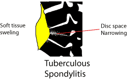
TUBERCULOSIS OF BONE

Tuberculosis affects primarily the lungs and gastrointestinal tract, but may also infect the musculoskeletal system.

Pathology

  • Hematogenous dissemination to long bones and vertebrae
  • Direct spread to bone from adjacent tuberculous lymphadenitis
  • Single or multi centric, particularly in AIDS
  • Predilection for synovium – higher oxygen pressure

There is caseating granulomatous inflammation with bone necrosis, resulting in the destruction of bone.

Extension of the TB osteomyelitis into joint spaces results in tuberculous arthritis. The infection crosses to the bone on the other side of the joint.

Incidence

  • 3% of tuberculosis is musculoskeletal. Of these, the commonest area affected is the spine.

Radiological signs

  • Lytic process.
  • Affects both sides of a joint or disc space.
  • Soft tissue swelling
    • Effusion in a joint
    • Paravertebral swelling in thorax
    • Fusiform paravertebral swelling in the neck
  • Cystic tuberculosis – causes cystic lesions in bones

Sclerosis is a sign of healing TB – successfully treated lesions become sclerotic as they heal.

Special Investigations

The ESR is raised often to above 100 mm/hr. On the differential white cell count there is a lymphocytosis. Sputum is taken for culture and a 24 hour urine collection can be taken if renal tuberculosis is a suspected cause.

Serological tests such as the Mantoux or Tine test are positive. In a third world population, such as South Africa there is anyway a high incidence of positivity with these tests. A positive test merely indicates that a patient has had exposure to TB in the past. A negative test may be of more value in excluding TB, but other immunosuppressive diseases such as HIV may suppress the reaction.

Biopsy

Biopsy may be needed is the diagnosis is in doubt. In South Africa, where tuberculosis is common, biopsy is not always done. If the blood and radiological criteria point to Tuberculosis therapy is started without a biopsy

Treatment

Anti tuberculous drugs are started.
(South African Medicines Formulary, fifth edition pg. 277)

Antituberculosis Drugs for Adults

Intensive phase (2 –3 months): Body weight <50kg >50kg
Rifampisin 120mg +Isoniasid (INH) 60 mg + Pirasinamide (PZA) 300mg + Etambutol 200mgCombination tablet (Rifafour®, Myrin Plus®) daily, 5 days per week 4 tablets daily 5 tablets daily
Maintenance phase: <50kg >50kg
Rifampisin 150mg + INH 100mgRifampisin 300mg + INH 150mg 3 tablets daily- -2 tablets daily

Pyridoxine (Vitamin B6) 10mg /day must be added in patients with inadequate intake, or at risk of developing peripheral neuropathy (pregnancy, diabetics, HIV infected patients)

Therapy lasts at least 1 year to 18 months. The ESR should show a decrease and the lesion on X rays becomes more sclerotic.

Failure to respond may be due to:-

  • Failure to take the drugs
  • Wrong diagnosis
  • Drug resistant tuberculosis

Side effects of anti tuberculous drugs

Serious: Drug causing Action
Jaundice (hepatitis) INH, Rifampisin, PZA Stop TB Drugs. Contact an infection specialist.
Skin rash with pyrexia INH, Rifampisin
Bleeding tenancy, shock, renal failure. Rifampisin
Impaired vision and color perception Etambutol

Splinting of joints

The affected joint must be immobiliased in a position of function. For the hip in the acute phase it means skin traction. Once discharged from hospital the patient with hip TB is told to sleep prone to avoid a flexion contracture. The spine will need an appropriate brace. For other joints a plaster cast, back slab or a custom made splint may be necessary

Place of Surgery

Surgery plays a limited role in the treatment of tuberculosis. Synovial biopsy may be needed to make the diagnosis. In spinal tuberculosis, gross instability or neurological fallout are indications for an anterior fusion and decompression.

Late surgery may be needed to repair the damage by a joint being destroyed or left in an unacceptable position. Arthrodesis is sometimes the only solution in these severe cases.

Spinal Tuberculosis

Thoracic spine in tuberculosis, showing disc narrowing and abscess

In the thoracic spine the patient presents with a gibbus.

There may be neurological fallout due to:

  • Mechanical pressure on the spinal cord
  • Vascular – blood supply to cord compromised
  • TB granuloma pressing on cord

Radiology

There is loss of disc space, soft tissue swelling is commonly seen adjacent to the involved area, or the pus may track into the psoas sheath (involvement about thoraco-lumbar junction)

Treatment principles remain the same as with TB of other areas. Antituberculosis medication is begun once the diagnosis is confirmed. Further deformity is prevented with a hard brace such as a Thoraco sacro lumbar orthosis (for Thoraco-lumbar involvement).

An indication for operation is neurological compromise, unresponsive to treatment. As TB spine involves mainly the vertebral body, an anterior approach is needed. The caseous material is curetted out and the vertebral bodies fused with an anterior bone graft. The rib removed for the thoracotomy can be used for this.

BONE TUMORS

Bone cancer, also known as primary bone cancer, is a form of cancer that develops in hard bone tissues and sometimes in the cartilage tissues of the bone. Primary bone cancer is rare, and differs from cancers that develop in: 1). other organs of the body and spread to the bones when metastasis (spreads), or 2). bone marrow cells (such as leukemia, multiplemyeloma, and lymphoma).

Neoplasms of the musculoskeletal system are of various types, including osteogenic, chondrogenic, fibrogenic, muscle (rhabdomyogenic), and marrow (reticulum) cell tumors as well as nerve, vascular, and fatty cell tumors. They may be primary tumors or metastatic tumors from primary cancers elsewhere in the body (eg, breast, lung, prostate, kidney). Metastatic bone tumors are more common than primary bone tumors.

Tumors may be benign (not cancer), or malignant (cancer). Also called neoplasm. Cancer is a term for diseases in which abnormal cells divide without control and can invade nearby tissues. Cancer cells can also spread to other parts of the body through the blood and lymph systems.

Types

Bone cancer is divided into separate types based on the type of cell where the cancer started. The most common type of bone cancer include:

  • Osteosarcoma. Osteosarcoma begins on bone cells. Osteosarcoma occurs most often in children and young adults.
  • Chondrosarcoma. Chondrosarcoma begins in the cartilage cells are normally found in the bone ends. Chondrosarcoma most commonly affects older adults.
  • Ewing’s Sarcoma. It is not clear where ewing sarcoma begins. Ewing’s sarcoma is believed that to begin in nerve tissue in the bone. Ewing’s sarcoma occurs most often in children and young adults.

Benign Bone Tumors

Benign bone tumors are slow growing, well circumscribed, and encapsulated. They produce few symptoms and do not cause death. Benign primary neoplasms of the musculoskeletal system include osteochondroma, enchondroma, bone cyst (eg, aneurysmal bone cyst), osteoid osteoma, rhabdomyoma, and fibroma. Benign tumors of the bone and soft tissue are more common than malignant primary bone tumors.

Osteochondroma, the most common benign bone tumor, may become malignant. Enchondroma is a common tumor of the hyaline cartilage of the hand, femur, tibia, or humerus. Osteoid osteoma is a painful tumor that occurs in children and young adults. Osteoclastomas (giant cell tumors) are benign for long periods but may invade local tissue and cause destruction. These tumors may undergo malignant transformation and metastasize. Bone cysts are expanding lesions within the bone (eg, aneurysmal and unicameral).

Malignant Bone Tumors

Primary malignant musculoskeletal tumors are relatively rare and arise from connective and supportive tissue cells (sarcomas) or bone marrow elements (myelomas). Malignant primary musculoskeletal tumors include osteosarcoma, chondrosarcoma, Ewing’s sarcoma, and fibrosarcoma. Soft tissue sarcomas include liposarcoma, fibrosarcoma, and rhabdomyosarcoma. Metastasis to the lungs is common. Osteogenic sarcoma (osteosarcoma) is the most common and is often fatal owing to metastasis to the lungs. It is seen most frequently in children, adolescents, and young adults (in bones that grow rapidly); in older people with Paget’s disease of the bone; and in persons with a prior history of radiation exposure. Common sites are distal femur, the proximal tibia, and the proximal humerus.

Chondrosarcoma, the second most common primary malignant bone tumor, is a large, bulky tumor that may grow and metastasize slowly or very fast, depending upon the characteristics of the tumor cells involved. Tumor sites may include pelvis, femur, humerus, spine, scapula, and tibia. Tumors may recur after treatment.

Metastatic Bone Disease

Metastatic bone disease (secondary bone tumors) is more common than any primary malignant bone tumor. The most common primary sites of tumors that metastasize to bone are the kidney, prostate, lung, breast, ovary, and thyroid. Metastatic tumors most frequently attack the skull, spine, pelvis, femur, and humerus and often involve more than one bone.

Stages of Bone Cancer

  1. Stage I: In this stage, the cancer is confined to the bone and hasn’t metastasized to other parts of the body. This stage is considered not aggressive and low grade.
  2. Stage II: In this stage, the cancer is confined to the bone and hasn’t metastasized to other parts of the body; however, biopsy testing reveals that the bone cancer is aggressive and high grade.
  3. Stage III: In this stage, the cancer is present in two or more different parts of the same bone. Biopsy testing reveals that the cancer can be either low grade or high grade.
  4. Stage IV: In this stage, the cancer has metastasized from the bone to other regions of the body, such as liver, lungs or brain.

Causes of Bone Cancer

The cause of many bone cancers is not clear. Doctors believe the cancer occurs due to an error in the DNA of a cell. This error causes the cell to grow and divide uncontrollably resulting in cancer. These abnormal cells continue to live instead of dying at a particular time. The buildup of these mutated cells result in a tumor/mass, which in turn infiltrates the nearby structures and/or metastasize to other parts of the body.

Risk Factors of Bone Cancer

  • Inherited genetic syndromes, such as hereditary retinoblastoma and Li-Fraumeni syndrome may cause or increase the risk of having bone cancer.
  • Precancerous conditions such as Paget’s disease of bone affects the older adults and also increases the risk of developing bone cancer.
  • Exposure to radiation from cancer treatment increases the risk of having a bone cancer in the future.

Symptoms of Bone Cancer

Characteristic feature of bone cancer or bone cancer symptoms include:
Bone pain.

  • Swelling and pain near the affected area.
  • Fracture.
  • Fatigue.
  • Weight loss is not desired.
  • Spinal metastasis results in cord compression and neurologic deficits (eg, progressive pain, weakness, gait abnormality, paresthesia, paraplegia, urinary retention, loss of bowel or bladder control).

Assessment and Diagnostic Findings

  • May be diagnosed incidentally after pathologic fracture
  • CT scan, bone scan, myelography, MRI, arteriography, x-ray studies
  • Biochemical assays of the blood and urine (alkaline phosphatase levels are frequently elevated with osteogenic sarcoma; serum acid phosphatase levels are elevated with metastatic carcinoma of the prostate; hypercalcemia is present with breast, lung, and kidney cancer bone metastases)
  • Surgical biopsy for histologic identification; staging based on tumor size, grade, location, and metastasis

Medical Management

The goal of treatment is to destroy or remove the tumor. This may be accomplished by surgical excision (ranging from local excision to amputation and disarticulation), radiation, or chemotherapy.

  • Limb-sparing (salvage) procedures are used to remove the tumor and adjacent tissue; surgical removal of the tumor may, however, require amputation of the affected extremity.
  • Chemotherapy is started before and continued after surgery in an effort to eradicate micrometastatic lesions.
  • Soft tissue sarcomas are treated with radiation, limb-sparing excision, and adjuvant chemotherapy.
  • Metastatic bone cancer treatment is palliative; therapeutic goal is to relieve pain and discomfort as much as possible while promoting quality of life.
  • Internal fixation of pathologic fractures, arthroplasty, or methylmethacrylate (bone cement) minimizes associated disability and pain in metastatic disease.

Nursing Interventions for Bone Cancer

Pain management

  • Psychological pain management techniques (deep breath relaxation techniques, visualization, and guided imagery) and pharmacological (providing analgesic).

Teach effective coping mechanisms

  • Motivation clients and families to express their feelings, and give moral support and encourage families to consult a psychologist or clergy.

Provide adequate nutrition

  • Decreased appetite, nausea, vomiting often occur as a side effect of chemotherapy and radiation, so it should be given adequate nutrition. Antiemetic and relaxation techniques can reduce gastrointestinal reactions. Parenteral nutrition can be carried out in accordance with the indications of the doctor.

Health education

  • Patients and families are given health education on the likelihood of complications, treatment programs, and wound care techniques at home .

If necessary; traction, Traction Treatment Principles

  • Provide comfort measures (eg frequently change position, back massage) and therapeutic activity.
  • Give the drug as an indication of examples; analgesic muscle relaxant.
  • Give local heating as indicated.
  • Give strength in early bandage / replacement in accordance with the indications, use aseptic technique correctly.
  • Keep linen remains dry, free of wrinkles.
  • Encourage the client to use loose cotton clothing.
  • Encourage the client to use stress management, for example: guided imagery, deep breathing.
  • Assess the degree of immobilization produced.
  • Identification signs or symptoms that require medical evaluation, eg edema, erythema.

Renal Failure:

Definition:

Kidney fails due to any dysfunction of normal renal function.

Types of Renal Failure:

Acute Renal Failure (ARF):

Definition:

It is an acute, severe, short timing duration of kidney disfunctioning in which kidney unable to remove the body metabolic waste products.

Etiology:

Pre_renal cause:

  1. Hemorrhage
  2. Loss of fluid
  3. Heart failure
  4. Myocardial infarction
  5. Anaphylactic shock
  6. Hypo_volumia Intra_renal cause:
  7. Renal ischemia
  8. Acute glomerulonephritis
  9. Pyelonephritis
  10. Kidney infection
  11. NSAIDS
  12. Heavy metal poison Post_renal cause:
  13. Renal tumor
  14. Benign, Prostate hyperplasia
  15. Ureter structure

Clinical features:

  1. Excessive Nitrogen in blood
  2. Dry skin
  3. Drowsiness
  4. Headache
  5. Seizures
  6. Blood in urine
  7. Creatinine level (elevated)
  8. Oliguria
  9. Hyperkalamia

Complications:

  1. Cardiac arrest
  2. Dysrhythmia
  3. Anemia
  4. Shock
  5. Hyperkalamia
  6. Dehydration
  7. Hiccough

Medical management

  1. diurectics
  2. tab:Lasix BD
  3. Chlorothiazide 500mg IV 8-12 hours
  4. Calcium carbonate in case of Hypo-calacimia
  5. Magnesium compound should not be given (Diarrhea)
  6. Aluminum chloride for correction of acidosis
  7. IV maintains
  8. ACE inhibitor

Investigations:

  1. KUB X-ray
  2. Urine DR
  3. CBC
  4. Urinanalysis
  5. CT scan
  6. MRI
  7. Renal ultrasound
  8. Pyelogram

Surgical Management:

  1. Dialysis
  2. Haemodylasis
  3. Peritoneal dialysis
  4. Renal replacement therapy

Nursing Management:

  1. Maintaining fluid & electrolyte balance
  2. Maintains of nutritional status
  3. Restricted for protein
  4. Maintain bed rest in acute case
  5. Prevent from injury, provide bed rails
  6. Avoid source of infection
  7. Identification of preventive environment or health factors contributing to the illness such as: hypertension & Nephrotoxic drugs.

2.. Chronic Renal Failure (CRF):

Definition:

Chronic Renal Failure involves progressive, irreversible destruction of the nephron in both kidneys. The disease process progress until most nephrons are destroyed & replaced by nonfunctional scar tissue.

Etiology:

Glomeruler dysfunction:

  1. Glomerulonephritis
  2. Diabetic Nephropathy
  3. Hypertensive Nephrosclerosis

Systemic disease:

  1. Sickle cell anemia
  2. Scleroderma
  3. Polyarteritis nodosa Urinary tract obstruction:
  4. Prostatic & bladder tumor
  5. Lymphodenopathy
  6. Calculi

Others:

  1. Chronic Pyelonephritis
  2. Nephritic Syndrome
  3. Renal infarction Clinical features:
  4. Hypertension
  5. Fatigue
  6. Pain
  7. Metabolic acidosis
  8. Pulmonary edema
  9. Anorexia
  10. Nausea
  11. Vomiting
  12. Diarrhea
  13. Lethargy
  14. Joint pain
  15. Pruitis Complication:
  16. CCF Investigation:
  17. ECG
  18. EEG
  19. BUN X-ray Medical Management:
  20. Anti-hypertensive drug
  21. Calcium Gluconate
  22. IV maintains Surgical Management:
  23. Dialysis

o Haemodylasis

o Peritoneal dialysis

o Renal replacement therapy Nursing Management:

  1. Maintaining fluids & electrolyte balance
  2. Maintain I/O chart
  3. Prevent infection or injury to promote skin care
  4. Promote comfort as medicate the patient as prescribed by the Doctor
  5. To keep lips moist, give oral hygiene
  6. Assist with coping in life style & self concept
  7. Avoidance of OTC (over-the-counter) drugs, specially aspirin, cold medication & NSAIDS.

Renal Calculi

Renal calculi are crystalline of minerals around the organic matrix such as: pus, blood or devitalized tissue. Most stones consist of calcium salts or magnesium-ammonium phosphate; the remainders are cystine or uric acid stones.

Etiology:

  1. Metabolic
  2. Diet
  3. Genetic factor
  4. Life style (Sedentary)
  5. Actual idiopathic
  6. Long term use of antacids, Vita-D, Vita-C

Clinical features:

  1. Abdominal pain
  2. Nausea
  3. Vomiting
  4. Colic pain
  5. Moist skin
  6. Urinary infection
  7. Fever
  8. Chills

Investigations:

  1. KUB X-ray
  2. Retrograde pyelography
  3. Ultrasound
  4. CT scan
  5. Cytoscopy
  6. Urinanalysis
  7. Serum Calcium level
  8. Serum Uric acid level

Medical Management:

  1. Analgesic
  2. Calcium oxalate
  3. Antimicrobial agents
  4. Surgical Management:
  5. Ureterolithotomy
  6. Pyelilithotomy
  7. Nephrolithotomy

Nursing Management:

  1. Increase for hydration
  2. Administer of the antimicrobial agents
  3. Reduce anxiety
  4. High fluid intake at least 3000ml per day.

Pneumonia (Pneumonitis)

Pneumonia is an inflammation of the lung parenchyma caused by various microorganisms, including bacteria, mycobacteria, fungi, and viruses. Pneumonitis is a more general term that describes an inflammatory process in the lung tissue that may predispose or place the patient at risk for microbial invasion.

Pathophysiology

Normally, the upper airway prevents potentially infectious particles from reaching the sterile lower respiratory tract. Pneumonia arises from normal flora present in patients whose resistance has been altered or from aspiration of flora present in the oropharynx; patients often have an acute or chronic underlying disease that impairs host defenses. Pneumonia may also result from bloodborne organisms that enter the pulmonary circulation and are trapped in the pulmonary capillary bed.

Pneumonia affects both ventilation and diffusion. An inflammatory reaction can occur in the alveoli, producing an exudate that interferes with the diffusion of oxygen and carbon dioxide. White blood cells, mostly neutrophils, also migrate into the alveoli and fill the normally air-filled spaces. Areas of the lung are not adequately ventilated because of secretions and mucosal edema that cause partial occlusion of the bronchi or alveoli, with a resultant decrease in alveolar oxygen tension. Bronchospasm may also occur in patients with reactive airway disease. Because of hypoventilation, a ventilation–perfusion mismatch occurs in the affected area of the lung. Venous blood entering the pulmonary circulation passes through the underventilated area and travels to the left side of the heart poorly oxygenated. The mixing of oxygenated and unoxygenated or poorly oxygenated blood eventually results in arterial hypoxemia.

If a substantial portion of one or more lobes is involved, the disease is referred to as lobar pneumonia. The term bronchopneumonia is used to describe pneumonia that is distributed in a patchy fashion, having originated in one or more localized areas within the bronchi and extending to the adjacent surrounding lung parenchyma. Bronchopneumonia is more common than lobar pneumonia.

Summary of Pathophysiology: Pneumonia is an infection of the lungs involving an acute inflammatory response that impairs the work of the alveoli and interferes with ventilation.

Classification
Pneumonia can be classified as:
• community-acquired pneumonia;
• hospital-acquired pneumonia;
• aspiration pneumonia;
• pneumonia in immunocompromised patients.
Clinical manifestations
The clinical manifestations of pneumonia are:

  • Shortness of breath due to inflammation within the lungs, impairing gas exchange
  • Difficulty breathing (dyspnea) due to inflammation and mucus within the lungs
  • Fever due to infectious process
  • Chills due to increased temperature
  • Cough due to mucous production and irritation of the airways
  • Crackles due to fluid within the alveolar space and smaller airways
  • Rhonchi due to mucus in airways; wheezing due to inflammation within the larger airways
  • Discolored, possibly blood-tinged, sputum due to irritation in the airways or microorganisms causing infection
  • Tachycardia and tachypnea as the body attempts to meet the demand for oxygen
  • Pain on respiration due to pleuritic inflammation, pleural effusion, or atelectasis development
  • Headache, muscle aches (myalgia), joint pains, or nausea may be present depending on the infecting organism

Diagnosis
The following investigations are used in diagnosis (Woodhead 2010):

  • Chest X-ray: Shadows on chest x-ray, indicating infiltration, may be in a lobar or segmental
    pattern or more scattered.
  • Blood analysis: Elevated WBC (leukocytosis) showing sign of infection.
  • Blood pressure monitoring;
  • Blood urea measurement;
  • Low oxygen saturation on pulse oximetry.
  • ABGs: Arterial blood gas may show low oxygen and elevated carbon dioxide levels.
  • Blood culture; Culture and sensitivity of the sputum to identify the infective agent and the
    appropriate antibiotics.
  • Sputum samples for C & S.

Assessment and management
Assessment of severity is important to determine the management of the patient with pneumonia, and the CURB65 score can be used for this. This scores 1 point for each of:

  • C (confusion);
  • U (blood urea >7 mmol/L);
  • R (respiratory rate >30/min);
  • B (blood pressure: systolic <90 mmHg, diastolic <60 mmHg);
  • 65 (>65 years of age).

Medical management includes the prompt and appropriate administration of intravenous antibiotic therapy, oxygen and intravenous fluids to correct the fluid balance. If an oxygen saturation of more than 92% is not achieved using oxygen, NIV may be considered.

  • Administer oxygen as needed.
  • For bacterial infections, administer antibiotics such as macrolides (azithromycin, clarithromycin), fluoroquinolones (levofloxacin, moxifloxacin), beta-lactams (amoxicillin/clavulanate, cefotaxime, ceftriaxone, cefuroxime axetil, cefpodoxime, ampicillin/sulbactam), or ketolide (telithromycin).
  • Administer antipyretics when fever >101 for patient comfort:
  • acetaminophen, ibuprofen
  • Administer brochodilators to keep airways open, enhance airflow if needed:
  • albuterol, metaproterenol, levalbuterol via nebulizer or metered dose inhaler
  • Increase fluid intake to help loosen secretions and prevent dehydration.
  • Instruct the patient on how to use the incentive spirometer to encourage deep breathing; monitor progress.

NURSING DIAGNOSES

  • Risk for aspiration
  • Impaired ventilation
  • Ineffective airway clearance

NURSING INTERVENTION

  • Monitor respiration for rate, effort, use of accessory muscles, skin color, and breath sounds.
  • Record fluid intake and output for differences, signs of dehydration.
  • Record sputum characteristics for changes in color, amount, and consistency.
  • Properly dispose of sputum.
  • Explain to the patient:
  • Take adequate fluids—3 liters per day—to prevent excess fluid loss through the respiratory system with exhalation.
  • Use of incentive spirometer.


Tuberculosis

Definition:

An infectious and communicable disease, acid fast, gram positive disease, it usually involves in the lungs but it also occurs in the any organ of the body.

Etiology:

  1. Mycobacterium Tuberculosis
  2. Clinical features:
  3. In early stage, asymptotic
  4. Fatigue
  5. Malaise
  6. Anorexia
  7. Weight loss
  8. Low grade fever
  9. Night sweats
  10. Chest pain
  11. Haemoptysis
  12. Chills

Investigations:

  1. Mantox test
  2. Chest X-ray
  3. CBC

Complications:

  1. Milliary tuberculosis
  2. Pleural effusion
  3. TB Pneumonia
  4. Respiratory failure
  5. Hypo-volumic shock
  6. Pleurisy

Medical Management:

  1. Bineekly treatment
o Isoniazid INH 15mg/kg
o Rifampicin 100mg/kg
o Streptomycin Inj: 25-30/kg
o Pyrazinamide 5-mg/kg

Nursing Management:

  1. To improve air-way maintains
  2. Inhalation should be given to liquefy sputum
  3. Sputum should be collected in early morning for Mantox test
  4. Patient should be isolated
  5. Advised for fluid intake & high protein diet as Carbohydrates

Patient should be encouraged for personal hygiene.

Chronic Obstructive Pulmonary Disorder (COPD)

Chronic Obstructive Pulmonary Disorder (COPD)

Chronic obstructive pulmonary disorder includes chronic Bronchitis, Bronchiectasis, Emphysema, Empyema and the Asthma.

Bronchitis

Definition:

Chronic bronchitis is defined by the presence of chronic productive cough for minimum of 3 months per year for at least consecutive years in patients in whom other causes have been excluded, it is characterized by physiologically by hyper-trophy & hyper secretion of bronchial mucous glands.

Etiology:

  1. Viral infection
  2. Bacterial infection
  3. Chemical irritants
  4. Clinical features:
  5. Cough
  6. Frequent respiratory infection
  7. Hypoxemia
  8. Hyperpnoea
  9. Cyanosis

Complications:

  1. Pulmonale
  2. Acute respiratory failure
  3. Peptic ulcer
  4. GERD
  5. Pneumonia

Investigations:

  1. X-ray film
  2. ABG’s
  3. ECG
  4. EEG
  5. Exercise testing with Oximetry

Medical Management:

  1. Medical therapy depends upon symptoms
  2. Specific therapy
  3. Bronchodilators
  4. Antibiotics
  5. Oxygen inhalation

Nursing Management:

  1. Promote patient’s comfort
  2. Prevent from further complication
  3. Provide ventilated rooms
  4. Promote secretion- removal
  5. Relaxation of exercise
  6. Oxygen inhalation

Bronchiectasis

Definition:

Bronchiectasis is a disorder characterized by permanent, abnormal or irreversible dilation of the bronchial tree or one or more large bronchi

Etiology:

  1. Bacteria
  2. Virus (Adeno-virus, Influenza)

Clinical features:

Sign:

  1. Cyanosis
  2. Cough Symptoms:
  3. Haemoptysis
  4. Dysponea
  5. Fatigue
  6. Weakness
  7. Weight loss

Investigations:

  1. X-ray chest
  2. Bronchography
  3. Sputum examination
  4. CT scanning

Medical Management:

  1. Antibiotics
  2. Bronchodilators

Surgical Management:

  1. Segmentectomy
  2. Lobectomy

Nursing Management:

  1. ROM of the Chest
  2. Maintaining good hydration to liquefy secretion
  3. Maintaining good hygiene including oral hygiene
  4. Adequate rest, diet, exercise
  5. Avoidance from smoking
  6. Provide ventilated room.

Emphysema

Definition:

Emphysema is defined as pathologically by destructive changes in alveolar walls and enlargement of air-spaces distal to the terminal non-respiratory bronchioles. It is characterized physiologically by increased lung compliance, decreased diffusing, capacity & increased air-way resistance

Etiology:

  1. Idiopathic
  2. Risk factors are:
  3. Smoking
  4. Heredity
  5. Clinical features:
  6. Dysponea
  7. Cough
  8. Hypoxemia
  9. Under weight
  10. Wheezing breath

Investigation:

  1. X-ray
  2. CBC

Medical Management:

  1. Oxygen therapy
  2. Physic-therapy

Nursing Management:

  1. ROM of the Chest
  2. Maintaining good hydration to liquefy secretion
  3. Maintaining good hygiene including oral hygiene
  4. Adequate rest, diet, exercise
  5. Avoidance from smoking
  6. Provide ventilated room

Pleurisy

Definition:

Inflammation of layers of the lungs (Pleura i.e. Parietal pleura/ visceral pleura).

Etiology:

  1. Tuberculosis
  2. Bronchitis
  3. Pleural infection
  4. Pneumonia
  5. Lung neoplasm

Clinical features:

  1. Chest pain
  2. Ribbing sound from sides of the lung
  3. Sharp knife like pain during inspiration
  4. Dysponea
  5. Coughing with pain

Investigations:

  1. X-ray chest
  2. CBC

Medical Management:

  1. Treat the causative agent
  2. Analgesic
  3. Antipyretic
  4. Antibiotic
  5. Expectorant
  6. Laxatives
  7. Oxygen inhalation

Nursing Management:

  1. ROM of the Chest
  2. Maintaining good hydration to liquefy secretion
  3. Maintaining good hygiene including oral hygiene
  4. Adequate rest, diet, exercise
  5. Avoidance from smoking
  6. Provide ventilated room
  7. Avoidance from the allergens

Emphysema

Definition:

Empysema is pus within a body cavity most often the pleural cavity.

Etiology:

  1. Pleural effusion
  2. Pneumonia
  3. Lung abscess
  4. Tuberculosis
  5. Fungal infection
  6. After thorax surgery
  7. Chest trauma

Clinical features:

  1. Cough
  2. Dysponea
  3. Tachyponea
  4. Tachycardia
  5. Fever
  6. Unilateral chest expansion
  7. Malaise
  8. Decrease appetite

Investigation:

  1. X-ray chest
  2. Thoracenthesis

Medical Management:

  1. Antibiotics Surgical Management:
  2. Thoracenthesis

Nursing Management:

  1. Oxygen inhalation
  2. Bed rest
  3. Deep breathing exercise
  4. Provide ventilated room
  5. Encouraging the patient for co-operation.